TEM of Skeletal Muscle and Basal Lamina
Numéro d’image : 12006978
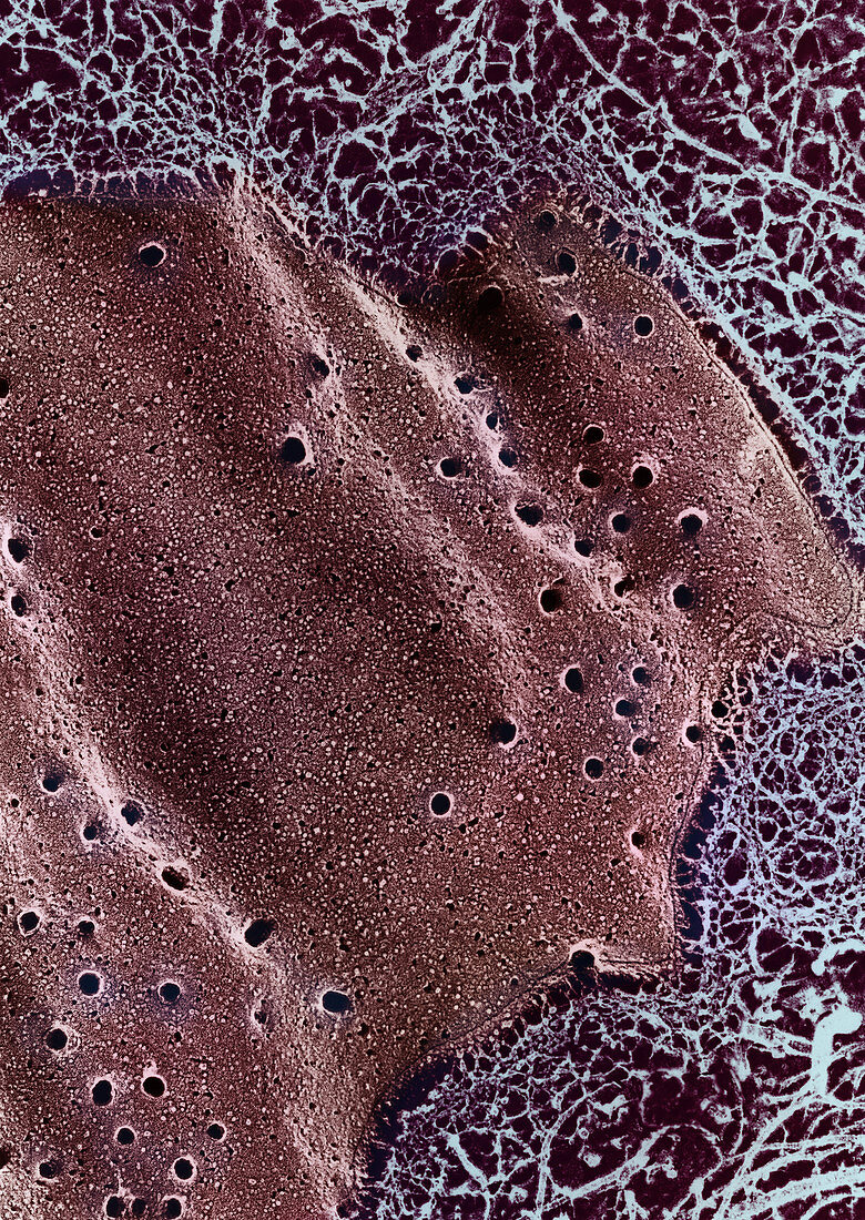
| Transmission electron micrograph of a quick-frozen,deeply-etched preparation of the sarcolemma of skeletal muscle and its basal lamina. The basal lamina are the fine mesh structures near the edge of the cell membrane. The larger fibrils at the edges of the image are collagen. Enhancement of 9C2723 | |
| Licence : | Droits gérés |
| Crédit: | Science Photo Library / Fawcett, Don W. |
| Taille de l’image : | 3552 px × 5010 px |
| Model Release : | Non requis |
| Property Release : | Non requis |
| Restrictions : |
|
Prix pour cette image À partir de 45 €
Produit vendu
(Calendrier, Carte postale, Carte de vœux, Impression sur textile, Packaging etc)
À partir de 45 €
Usage commercial
(Affichage, Annonce presse, Annonce TV, Carte, Digital - hors rés. sociaux, Digital - rés. sociaux etc)
À partir de 45 €
Éditorial
(Digital, Journal, Livre, Livre pratique, Magazine, Télévision etc)
À partir de 60 €
Usage non-commercial
(Digital - hors rés. sociaux, Digital - rés. sociaux etc)
À partir de 120 €
Mots clés
- basal lamina,
- cellule,
- collagène,
- cryo-fixation,
- cryofixation,
- cryofixé,
- cryofracture,
- fibrillaire,
- fibrille,
- fracture de gel,
- fracture due au gel,
- frcturé par le froid,
- histologie,
- M.E.T.,
- membrane,
- MET,
- micrographie,
- micrographie électronique à transmission,
- microscope,
- microscope électronique à transmission,
- microscope électronique en transmission,
- microscopie,
- muscle squelettique,
- rupture par congélation,
- sarcolemme