Oligodendroglioma
Numéro d’image : 12006772
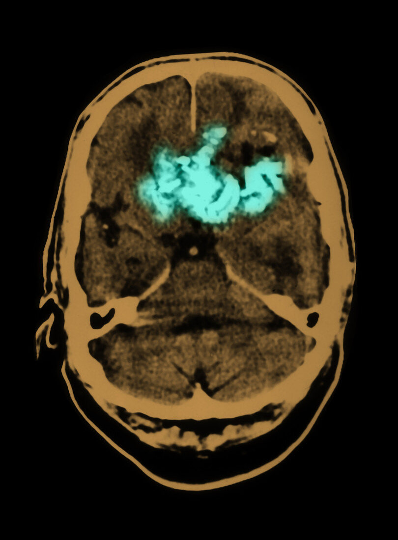
| This axial (cross sectional) CT image of the brain show a large,prominently calcified,heterogenous frontal lobe mass (shown in light green) with associated mass effect. There is surrounding edema.This is a primary brain tumor called an oligodendroglioma.This is a type of primary intracranial tumor called a glioma which arise from the supporting elements. In this case the oligodendrocytes are glial cells which normally are involved with the formation of myelin. These types of tumors have a very high incidence of calcifications. This is a colorized version of BF3177 | |
| Licence : | Droits gérés |
| Crédit: | Science Photo Library / Medical Body Scans |
| Taille de l’image : | 5395 px × 7324 px |
| Model Release : | Non requis |
| Property Release : | Non requis |
| Restrictions : |
|
Prix pour cette image À partir de 45 €
Produit vendu
(Calendrier, Carte postale, Carte de vœux, Impression sur textile, Packaging etc)
À partir de 45 €
Usage commercial
(Affichage, Annonce presse, Annonce TV, Carte, Digital - hors rés. sociaux, Digital - rés. sociaux etc)
À partir de 45 €
Éditorial
(Digital, Journal, Livre, Livre pratique, Magazine, Télévision etc)
À partir de 60 €
Usage non-commercial
(Digital - hors rés. sociaux, Digital - rés. sociaux etc)
À partir de 120 €