Active Demyelination of Spinal Cord in MS
Numéro d’image : 12006639
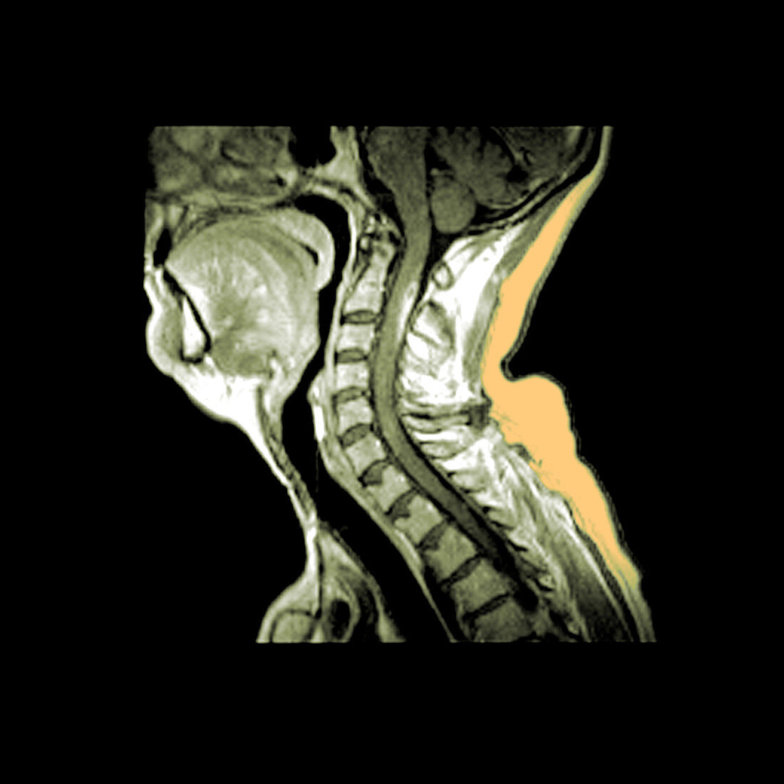
| This contrast enhanced sagittal T1 weighted MRI sequence of the cervical spine/spinal cord shows an area of pathologic enhancement along the posterior (dorsal) aspect of the upper cervical spinal cord. This is in a patient with multiple sclerosis and represents areas of active demyelination. Color-enhanced version of BG3562 | |
| Licence : | Droits gérés |
| Crédit: | Science Photo Library / Medical Body Scans |
| Taille de l’image : | 3600 px × 3600 px |
| Model Release : | Non requis |
| Property Release : | Non requis |
| Restrictions : |
|
Prix pour cette image À partir de 45 €
Produit vendu
(Calendrier, Carte postale, Carte de vœux, Impression sur textile, Packaging etc)
À partir de 45 €
Usage commercial
(Affichage, Annonce presse, Annonce TV, Carte, Digital - hors rés. sociaux, Digital - rés. sociaux etc)
À partir de 45 €
Éditorial
(Digital, Journal, Livre, Livre pratique, Magazine, Télévision etc)
À partir de 60 €
Usage non-commercial
(Digital - hors rés. sociaux, Digital - rés. sociaux etc)
À partir de 120 €
Mots clés
- anormal,
- augmenter,
- couleur,
- démyélinisation,
- I.R.M.,
- image de résonance magnétique,
- imagerie par résonnance magnétique,
- IRM,
- m.s.,
- maladie,
- médical,
- médicale,
- milliseconde,
- moelle épinière,
- ms,
- pathologie,
- rachis cervical,
- sagittal,
- sagittale,
- sclérose en plaques,
- scléroses,
- sclerosis,
- système nerveux central