Cerebellar Infarcts on MRI
Numéro d’image : 12006574
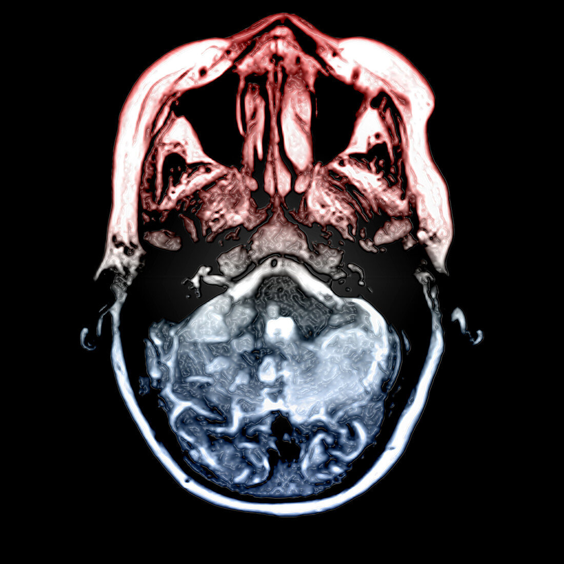
| This color enhanced axial (cross sectional) T2 weighted MRI image of the brain demonstrates multiple bilateral cerebellar infarctions (strokes) which are the focal regions of whiter density in the blue lower part of the image. There is also a small infarct (stroke) involving the posterior aspect of the brainstem (pons) on the right (your left) | |
| Licence : | Droits gérés |
| Crédit: | Science Photo Library / Living Art Enterprises |
| Taille de l’image : | 3600 px × 3600 px |
| Model Release : | Non requis |
| Property Release : | Non requis |
| Restrictions : |
|
Prix pour cette image À partir de 45 €
Produit vendu
(Calendrier, Carte postale, Carte de vœux, Impression sur textile, Packaging etc)
À partir de 45 €
Usage commercial
(Affichage, Annonce presse, Annonce TV, Carte, Digital - hors rés. sociaux, Digital - rés. sociaux etc)
À partir de 45 €
Éditorial
(Digital, Journal, Livre, Livre pratique, Magazine, Télévision etc)
À partir de 60 €
Usage non-commercial
(Digital - hors rés. sociaux, Digital - rés. sociaux etc)
À partir de 120 €