Dyke-Davidoff Masson Syndrome
Numéro d’image : 12006544
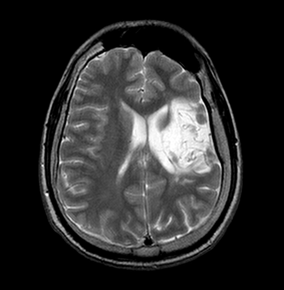
| This axial (cross sectional) MR image of the brain demonstrates an area of infarction (large white region on your right). This loss of blood supply occurred prenatally in the left cerebral hemisphere (right side of image) with subsequent hemiatrophy (looks smaller) of the same hemisphere,thickening of the left calvarium (on your right) and enlargement of the left frontal sinus. This constellation of findings represents the Dyke-Davidoff Masson syndrome | |
| Licence : | Droits gérés |
| Crédit: | Science Photo Library / Living Art Enterprises |
| Taille de l’image : | 3600 px × 3675 px |
| Model Release : | Non requis |
| Property Release : | Non requis |
| Restrictions : |
|
Prix pour cette image À partir de 45 €
Produit vendu
(Calendrier, Carte postale, Carte de vœux, Impression sur textile, Packaging etc)
À partir de 45 €
Usage commercial
(Affichage, Annonce presse, Annonce TV, Carte, Digital - hors rés. sociaux, Digital - rés. sociaux etc)
À partir de 45 €
Éditorial
(Digital, Journal, Livre, Livre pratique, Magazine, Télévision etc)
À partir de 60 €
Usage non-commercial
(Digital - hors rés. sociaux, Digital - rés. sociaux etc)
À partir de 120 €