Arachnoid Cyst
Numéro d’image : 12006511
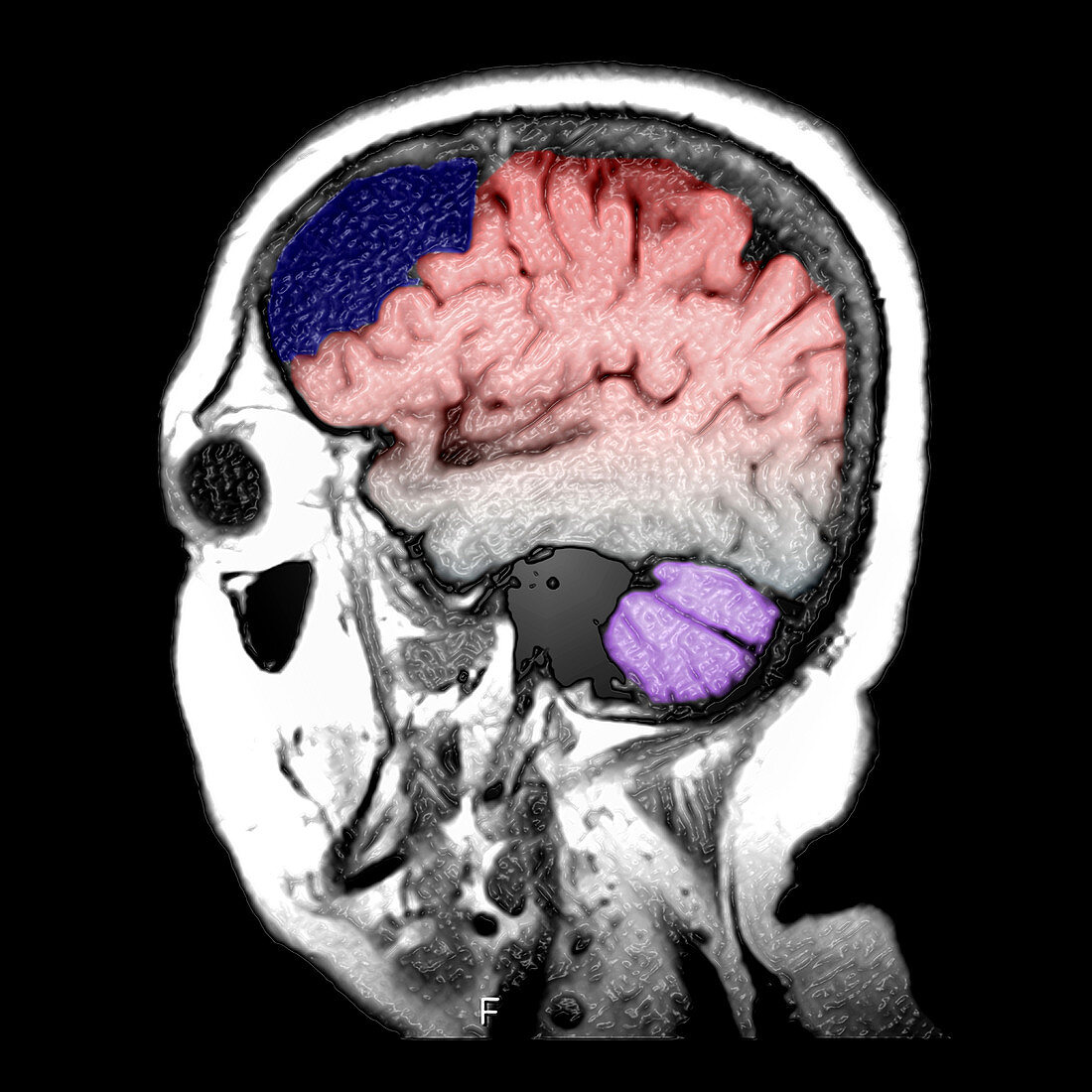
| This color enhanced sagittal (from the side) T1 weighted MRI image of the brain demonstrates an extra-axial collection of fluid,identical to CSF with associated mass effect upon the frontal lobe which represents a benign condition called an arachnoid cyst which is blue. The cerebellum is purple. The frontal and parietal lobes are red | |
| Licence : | Droits gérés |
| Crédit: | Science Photo Library / Living Art Enterprises |
| Taille de l’image : | 3600 px × 3600 px |
| Model Release : | Non requis |
| Property Release : | Non requis |
| Restrictions : |
|
Prix pour cette image À partir de 45 €
Produit vendu
(Calendrier, Carte postale, Carte de vœux, Impression sur textile, Packaging etc)
À partir de 45 €
Usage commercial
(Affichage, Annonce presse, Annonce TV, Carte, Digital - hors rés. sociaux, Digital - rés. sociaux etc)
À partir de 45 €
Éditorial
(Digital, Journal, Livre, Livre pratique, Magazine, Télévision etc)
À partir de 60 €
Usage non-commercial
(Digital - hors rés. sociaux, Digital - rés. sociaux etc)
À partir de 120 €
Mots clés
- arachnoiïde,
- bénin,
- cerveau,
- côté,
- couleur,
- coupe transversale,
- crâne,
- cranium,
- fluide cérébrospinal,
- fosse crânienne,
- humain,
- I.R.M.,
- image de résonance magnétique,
- imagerie par résonance magnétique,
- imagerie par résonnance magnétique,
- IRM,
- kyste,
- kyste arachnoïdien,
- liquide céphalo-rachidien,
- liquide céphalorachidien,
- médical,
- médicale,
- neuro-imagerie,
- neuroimagerie,
- neurologie,
- personne,
- sagittal,
- sagittale,
- scanner du cerveau,
- science