MRI of Stroke
Numéro d’image : 12003886
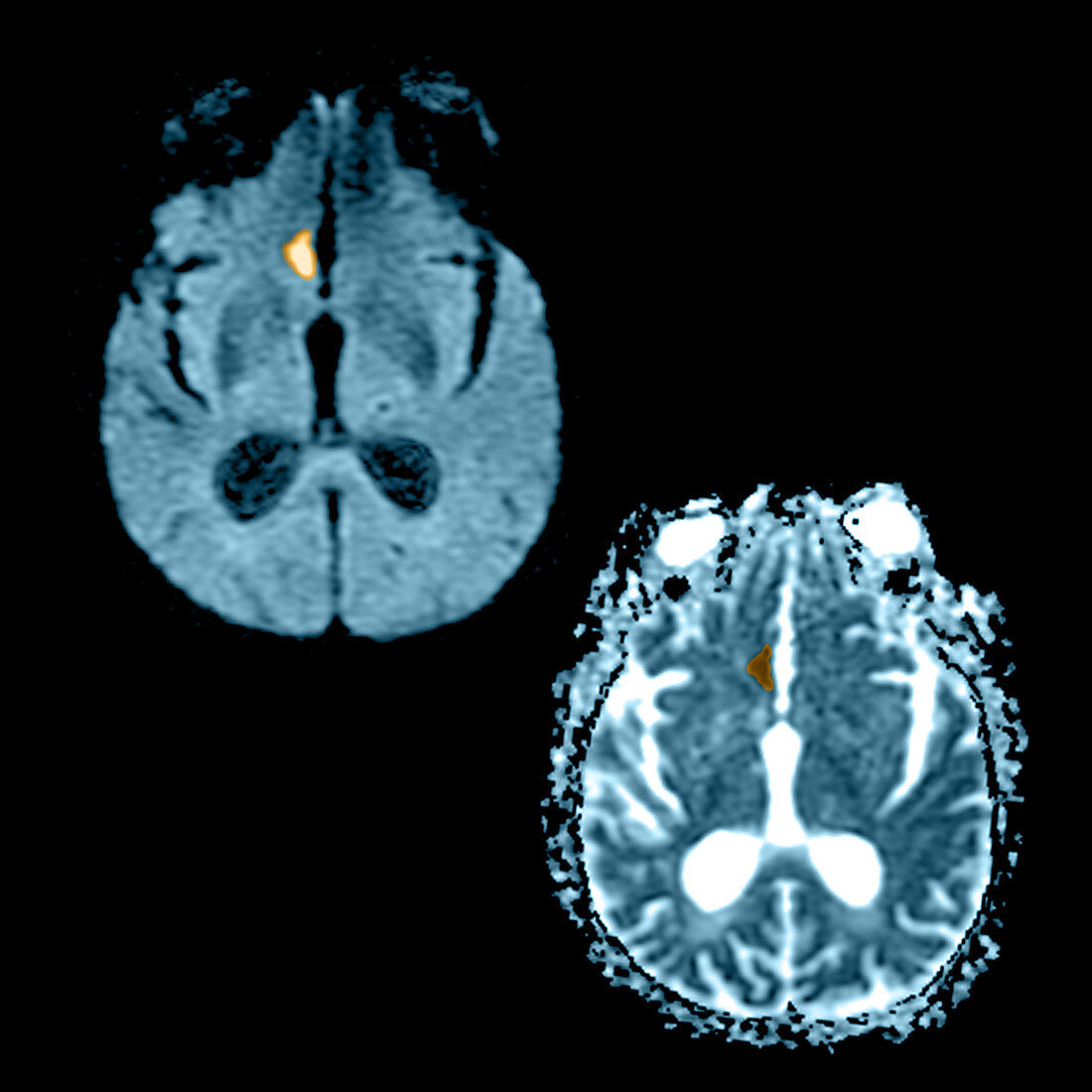
| This composite of 2 axial MRI images reveal the appearance of an acute stroke on diffusion weighted images and the corresponding ADC (apparrent diffusion coefficient) map. The diffusion weighted image in the upper left hand side shows a focal region of increased signal (looks white) in the inferior-medial frontal lobe. The ADC map in the lower right hand side shows that there is restricted (looks dark) diffusion in this region which indicates that the abnormality most likely represents an acute (usually looks like this for 10-14 days) infarct | |
| Licence : | Droits gérés |
| Crédit: | Science Photo Library / Medical Body Scans |
| Taille de l’image : | 4800 px × 4800 px |
| Model Release : | Non requis |
| Property Release : | Non requis |
| Restrictions : |
|
Prix pour cette image À partir de 45 €
Produit vendu
(Calendrier, Carte postale, Carte de vœux, Impression sur textile, Packaging etc)
À partir de 45 €
Usage commercial
(Affichage, Annonce presse, Annonce TV, Carte, Digital - hors rés. sociaux, Digital - rés. sociaux etc)
À partir de 45 €
Éditorial
(Digital, Journal, Livre, Livre pratique, Magazine, Télévision etc)
À partir de 60 €
Usage non-commercial
(Digital - hors rés. sociaux, Digital - rés. sociaux etc)
À partir de 120 €