Embryo at 26 days
Numéro d’image : 11999829
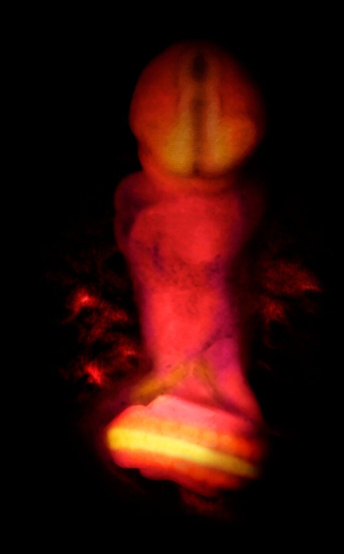
| Embryo at 26 days,computer-generated image from a micro-MRI scan. At this stage the embryo measures 4 millimetres in length. The head region (top) is bent downwards so that the developing central nervous system can be seen. The tube-like structure in the head region indicates the developing spinal cord. The tail of the embryo can be seen curving upwards so that the spinal cord and somites (muscle blocks,orange) are seen | |
| Licence : | Droits gérés |
| Crédit: | Science Photo Library / Anatomical Travelogue |
| Taille de l’image : | 3319 px × 5334 px |
| Model Release : | Non requis |
| Property Release : | Non requis |
| Restrictions : |
|
Prix pour cette image À partir de 45 €
Produit vendu
(Calendrier, Carte postale, Carte de vœux, Impression sur textile, Packaging etc)
À partir de 45 €
Usage commercial
(Affichage, Annonce presse, Annonce TV, Carte, Digital - hors rés. sociaux, Digital - rés. sociaux etc)
À partir de 45 €
Éditorial
(Digital, Journal, Livre, Livre pratique, Magazine, Télévision etc)
À partir de 60 €
Usage non-commercial
(Digital - hors rés. sociaux, Digital - rés. sociaux etc)
À partir de 120 €
Mots clés
- anatomie,
- anatomique,
- biologie,
- biologique,
- blocs,
- cerveau,
- développement,
- développement embryonnaire,
- embryologie,
- embryon,
- embryonnaire,
- foetal,
- foetale,
- fœtale,
- foetus,
- frontal,
- généré par ordinateur,
- humain,
- I.R.M.,
- imagerie par résonnance magnétique,
- IRM,
- micro irm,
- moelle épinière,
- organe,
- organes,
- personne,
- queue,
- reproduction,
- seul,
- somites,
- système nerveux central,
- unique