Breathing tube of fruit fly pupa
Numéro d’image : 11908383
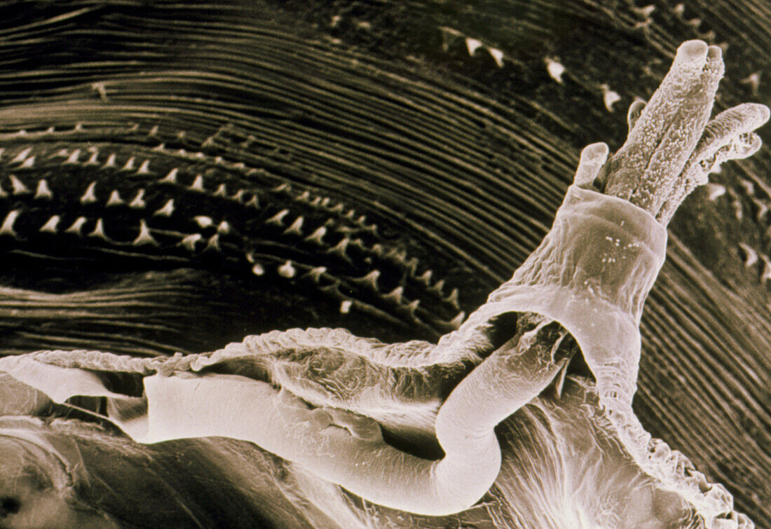
| Scanning electron micrograph of an anterior spiracle,or breathing tube,one of a pair,on the shed pupal casing of a fruit fly Drosophila melanogaster (wild type Oregon R). They project outward from the puparium,the outer grooved surface of which is seen in the background. This anterior spiracle terminates in tubelike extensions,called spiracular papillae (seen here). The tips of these open in narrow slits,which take in air & pass it to the developing pupa. They air is circulated through the thick tracheal tube or pipe lying on the inner surface of the puparium. Magnification: x450 at 10x8 inch,x56 at 35mm size | |
| Licence : | Droits gérés |
| Crédit: | Science Photo Library / Burgess, Dr. Jeremy |
| Taille de l’image : | 3614 px × 2480 px |
| Model Release : | Non requis |
| Property Release : | Non requis |
| Restrictions : | - |
Prix pour cette image À partir de 45 €
Produit vendu
(Calendrier, Carte postale, Carte de vœux, Impression sur textile, Packaging etc)
À partir de 45 €
Usage commercial
(Affichage, Annonce presse, Annonce TV, Carte, Digital - hors rés. sociaux, Digital - rés. sociaux etc)
À partir de 45 €
Éditorial
(Digital, Journal, Livre, Livre pratique, Magazine, Télévision etc)
À partir de 60 €
Usage non-commercial
(Digital - hors rés. sociaux, Digital - rés. sociaux etc)
À partir de 120 €