First whole body X-ray,1897
Numéro d’image : 11905359
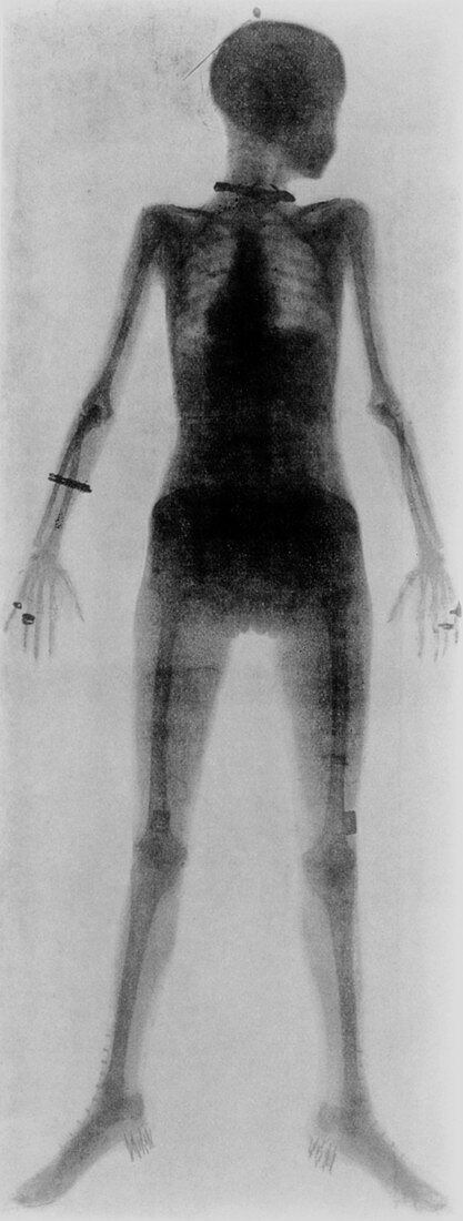
| First whole body X-ray (1907). The first whole body radiograph ever taken of a living person,a woman in 1897. The X-ray was made from a single exposure by William Morton of New York. Standing with her head in profile,her skeleton,heart and liver are seen. Her jewellery is highly visible: hatpin,necklace,bracelet,rings. On her feet she is wearing high button boots with nailed-on heels,and around her hips and abdomen a whalebone corset. To make this X-ray,Morton included a 12 inch induction coil with the current supplied from a New York street mains connection. A Crookes' tube was positioned 54 inches from the X-ray plate and the time taken was about 30 minutes | |
| Licence : | Droits gérés |
| Crédit: | Science Photo Library |
| Taille de l’image : | 1863 px × 4919 px |
| Model Release : | Non requis |
| Property Release : | Non requis |
| Restrictions : | - |
Prix pour cette image À partir de 45 €
Produit vendu
(Calendrier, Carte postale, Carte de vœux, Impression sur textile, Packaging etc)
À partir de 45 €
Usage commercial
(Affichage, Annonce presse, Annonce TV, Carte, Digital - hors rés. sociaux, Digital - rés. sociaux etc)
À partir de 45 €
Éditorial
(Digital, Journal, Livre, Livre pratique, Magazine, Télévision etc)
À partir de 60 €
Usage non-commercial
(Digital - hors rés. sociaux, Digital - rés. sociaux etc)
À partir de 120 €