Whole body scans
Numéro d’image : 11877102
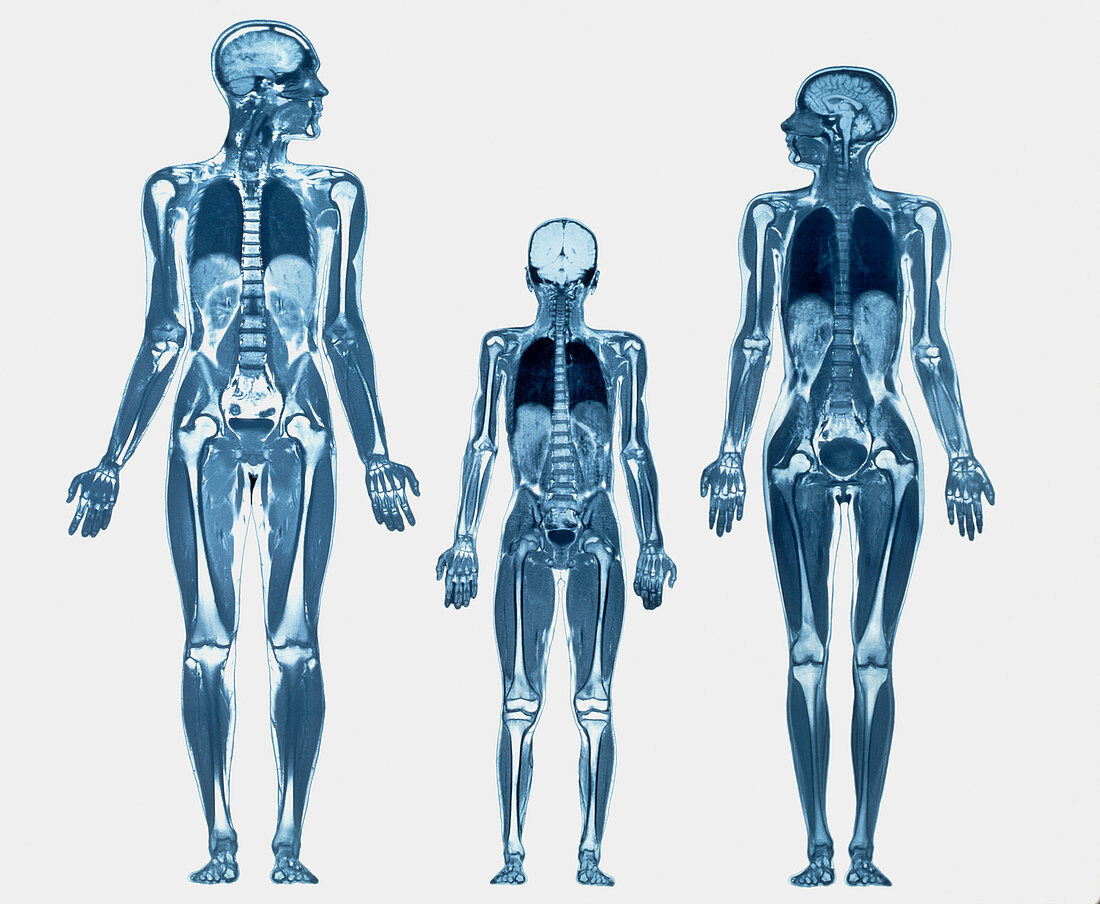
| Whole body scans. Coloured magnetic resonance imaging (MRI) whole body scans of a man (left),a woman and a nine year old boy in coronal (frontal) section. The whitish skeletons consist of the long bones of the limbs and the vertebrae of the spine. At top the brains are seen in their skulls. The lungs (dark) in their chests,the lobes of the livers (pale ovals) in their abdomens and the bladders (dark ovals) in their pelvises are also visible. These whole body images are composites of many MRI scans made along the length of the body and then combined. MRI scanning uses radio waves and a powerful magnetic field to produce slice images through the body | |
| Licence : | Droits gérés |
| Crédit: | Science Photo Library / Fraser, Simon |
| Taille de l’image : | 4724 px × 3884 px |
| Model Release : | Non requis |
| Property Release : | Non requis |
| Restrictions : | - |
Prix pour cette image À partir de 45 €
Produit vendu
(Calendrier, Carte postale, Carte de vœux, Impression sur textile, Packaging etc)
À partir de 45 €
Usage commercial
(Affichage, Annonce presse, Annonce TV, Carte, Digital - hors rés. sociaux, Digital - rés. sociaux etc)
À partir de 45 €
Éditorial
(Digital, Journal, Livre, Livre pratique, Magazine, Télévision etc)
À partir de 60 €
Usage non-commercial
(Digital - hors rés. sociaux, Digital - rés. sociaux etc)
À partir de 120 €
Mots clés
- anatomie,
- corps humain,
- diagnostic,
- diagnostics,
- diagnostique,
- diagnostiques,
- en bonne santé,
- enfant,
- famille,
- femme,
- homme,
- I.R.M.,
- imagerie par résonance magnétique,
- imagerie par résonnance magnétique,
- IRM,
- médecine,
- médical,
- médicale,
- résonance magnétique nucléaire,
- RMN,
- sain,
- soins de santé,
- squelette