Col. MRI scan of thorax & abdomen of elderly woman
Numéro d’image : 11877074
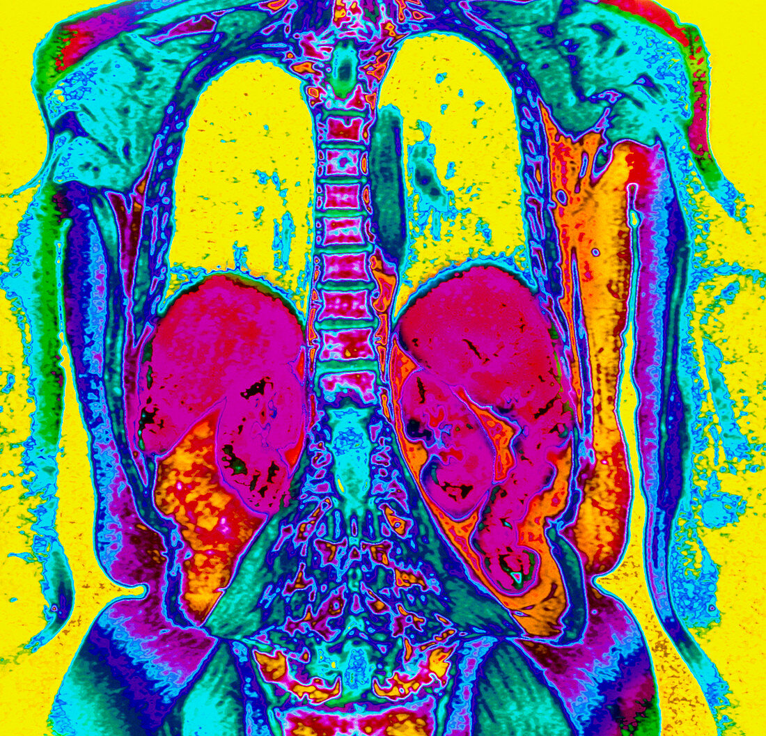
| Thorax and abdomen. Coloured Magnetic Resonance Imaging (MRI) scan of the thorax and abdomen of a woman aged 58 years,seen in posterior view. The arms are seen on either side,with the waist and hips at lower frame. The thorax contains the yellow lung fields (upper frame) with bones of the thoracic spine at upper centre. In the abdomen are lobes of the liver (pink,below the lungs) and a pair of kidneys (pink,below the liver lobes). Bands of muscles can be seen around the lumbar spine (lower centre) and the shoulders (at top,green-blue). MRI scanning creates "slice" images through the body using radio waves | |
| Licence : | Droits gérés |
| Crédit: | Science Photo Library / Fraser, Simon / Newcastle Upon Tyne / Royal Victoria Infirmary |
| Taille de l’image : | 4625 px × 4446 px |
| Model Release : | Non requis |
| Property Release : | Non requis |
| Restrictions : | - |
Prix pour cette image À partir de 45 €
Produit vendu
(Calendrier, Carte postale, Carte de vœux, Impression sur textile, Packaging etc)
À partir de 45 €
Usage commercial
(Affichage, Annonce presse, Annonce TV, Carte, Digital - hors rés. sociaux, Digital - rés. sociaux etc)
À partir de 45 €
Éditorial
(Digital, Journal, Livre, Livre pratique, Magazine, Télévision etc)
À partir de 60 €
Usage non-commercial
(Digital - hors rés. sociaux, Digital - rés. sociaux etc)
À partir de 120 €