Normal bone scan of chest: posterior view
Numéro d’image : 11877058
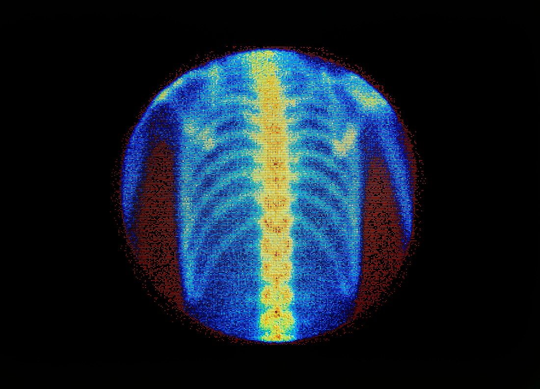
| False-colour scintigram (bone scan) of a normal chest in posterior view. The scintigram is a map of radioactivity from a tracer,which is injected into the subject prior to taking the scan & which becomes concentrated in bone. Radioactivity passes through the body to be recorded by an external gamma camera. Technetium-99m linked to MDP is the tracer of choice for bone scans. This technique is used to screen cancer patients for secondary disease (metastases) in the follow-up to treatment of their primary cancer. The increased uptake of radioactivity in cancerous bone is revealed as abnormal,"hot spots" on a bone scan | |
| Licence : | Droits gérés |
| Crédit: | Science Photo Library / MEDICAL PHYSICS, RVI, NEWCASTLE UPON-TYNE / SIMON FRASER |
| Taille de l’image : | 4935 px × 3555 px |
| Model Release : | Non requis |
| Property Release : | Non requis |
| Restrictions : | - |
Prix pour cette image À partir de 45 €
Produit vendu
(Calendrier, Carte postale, Carte de vœux, Impression sur textile, Packaging etc)
À partir de 45 €
Usage commercial
(Affichage, Annonce presse, Annonce TV, Carte, Digital - hors rés. sociaux, Digital - rés. sociaux etc)
À partir de 45 €
Éditorial
(Digital, Journal, Livre, Livre pratique, Magazine, Télévision etc)
À partir de 60 €
Usage non-commercial
(Digital - hors rés. sociaux, Digital - rés. sociaux etc)
À partir de 120 €