False-colour bone scintigram of a healthy adult
Numéro d’image : 11877054
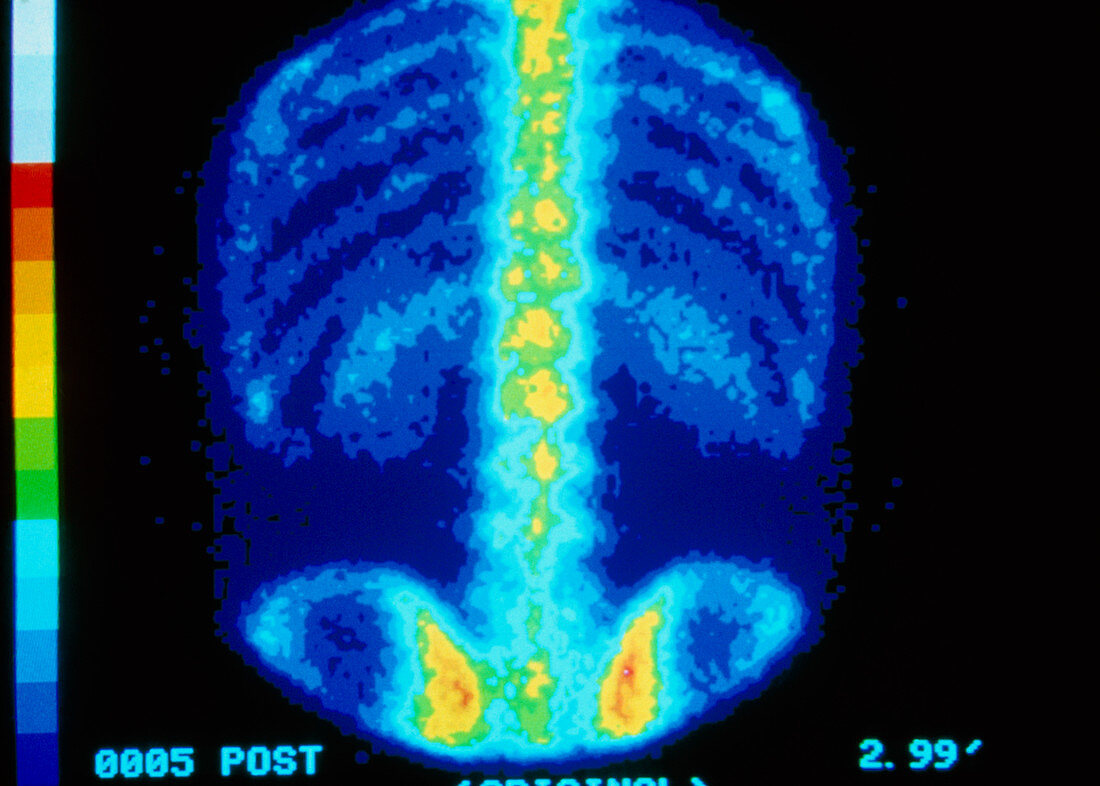
| False colour bone scintigram (gamma camera scan) of a coronal (frontal) view of a normal adult skeleton,showing ribs,thoracic & lumbar spine and pelvis (bottom). Bone scans record the distribution & intensity of gamma radiations emanating from a radionuclide injected into the body prior to taking the scan,which concentrates in bone. A crystal scintillator in the camera resolves radiations as flashes of light that are detected by a system of photomultipliers. Bone scans are used to assess presence & extent of bone cancers,which present as sites of increased radionuclide uptake that appear as brighter,"hot spots" on the image | |
| Licence : | Droits gérés |
| Crédit: | Science Photo Library / CNRI |
| Taille de l’image : | 3873 px × 2769 px |
| Model Release : | Non requis |
| Property Release : | Non requis |
| Restrictions : | - |
Prix pour cette image À partir de 45 €
Produit vendu
(Calendrier, Carte postale, Carte de vœux, Impression sur textile, Packaging etc)
À partir de 45 €
Usage commercial
(Affichage, Annonce presse, Annonce TV, Carte, Digital - hors rés. sociaux, Digital - rés. sociaux etc)
À partir de 45 €
Éditorial
(Digital, Journal, Livre, Livre pratique, Magazine, Télévision etc)
À partir de 60 €
Usage non-commercial
(Digital - hors rés. sociaux, Digital - rés. sociaux etc)
À partir de 120 €