Coloured SEM of secretory cells in adrenal gland
Numéro d’image : 11876698
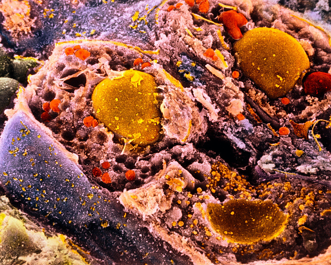
| Adrenal gland cells. Coloured Scanning Electron Micrograph (SEM) of secretory cells of the zona fasciculata region of the adrenal gland. Three cells are seen,sectioned by cell fracture to show internal anatomy. There is a large rounded nucleus (brown) in two of the cells. The cell cytoplasm is filled with lipid droplets (orange); larger mitochondria (red) are seen at upper right; the cell membrane is coloured blue. Secretory cells in this part of the adrenal cortex produce glucocorticoid hormones such as cortisol. These hormones have wide-ranging metabolic effects on the body. Magnification: x6,100 at 6x7cm size. x8,700 at 4x5ins | |
| Licence : | Droits gérés |
| Crédit: | Science Photo Library / UNIVERSITY LA SAPIENZA, ROME / DEPT. OF ANATOMY / PROF. P. MOTTA |
| Taille de l’image : | 4561 px × 3645 px |
| Model Release : | Non requis |
| Property Release : | Non requis |
| Restrictions : | - |
Prix pour cette image À partir de 45 €
Produit vendu
(Calendrier, Carte postale, Carte de vœux, Impression sur textile, Packaging etc)
À partir de 45 €
Usage commercial
(Affichage, Annonce presse, Annonce TV, Carte, Digital - hors rés. sociaux, Digital - rés. sociaux etc)
À partir de 45 €
Éditorial
(Digital, Journal, Livre, Livre pratique, Magazine, Télévision etc)
À partir de 60 €
Usage non-commercial
(Digital - hors rés. sociaux, Digital - rés. sociaux etc)
À partir de 120 €