Skin layers,SEM
Numéro d’image : 11876316
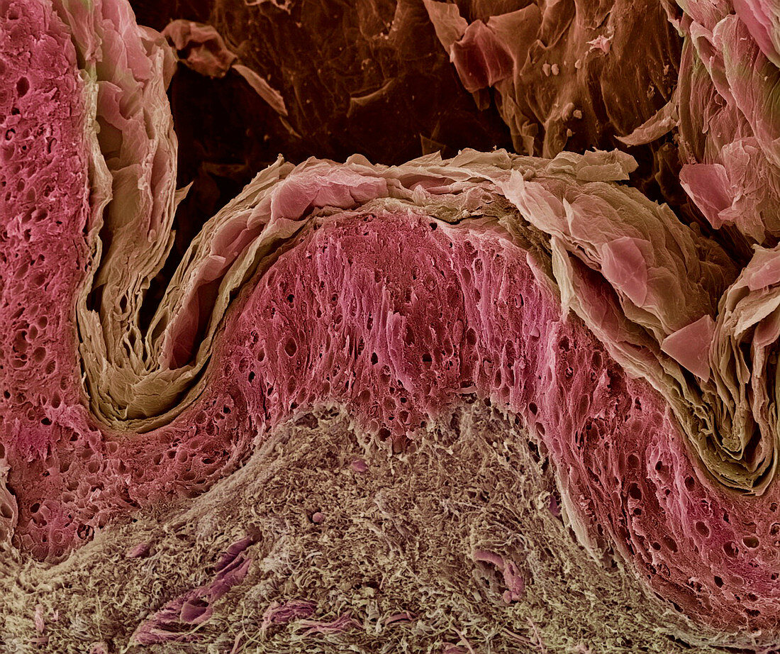
| Skin. Coloured scanning electron micrograph (SEM) of freeze-fractured human skin. The top layer is the stratum corneum (flaky,pale brown),a cornified layer of the epidermis that is composed of flattened,dead skin cells that form the surface of the skin. The dead cells from this layer are continuously being shed and replaced by cells from the living epidermal layer below (pink). The lowest layer seen here is the dermis (grey-brown,lower centre). This is a thick layer of fibrous connective tissue that supports and nourishes the epidermis. The skin is the body's largest organ,accounting for around 15% of the body's weight. Magnification: x220 at 6x7cm size | |
| Licence : | Droits gérés |
| Crédit: | Science Photo Library / Gschmeissner, Steve |
| Taille de l’image : | 3543 px × 2970 px |
| Model Release : | Non requis |
| Property Release : | Non requis |
| Restrictions : | - |
Prix pour cette image À partir de 45 €
Produit vendu
(Calendrier, Carte postale, Carte de vœux, Impression sur textile, Packaging etc)
À partir de 45 €
Usage commercial
(Affichage, Annonce presse, Annonce TV, Carte, Digital - hors rés. sociaux, Digital - rés. sociaux etc)
À partir de 45 €
Éditorial
(Digital, Journal, Livre, Livre pratique, Magazine, Télévision etc)
À partir de 60 €
Usage non-commercial
(Digital - hors rés. sociaux, Digital - rés. sociaux etc)
À partir de 120 €
Mots clés
- agrandissement,
- anatomie,
- avion,
- catégorie,
- cellule,
- cellules,
- coloré,
- colorié,
- colorisé,
- corps humain,
- couche cornée,
- couches,
- coupe,
- coupe transversale,
- cryofracture,
- derme,
- dermis,
- divisé,
- épiderme,
- fracture,
- fracturé,
- fracture de gel,
- fracture due au gel,
- histologie,
- images,
- M.E.B.,
- MEB,
- microscope électronique à balayage,
- mort,
- partie,
- peau,
- photos au microscope,
- section,
- stratum corneum,
- sujets,
- surface,
- tissus