Skin section,SEM
Numéro d’image : 11876292
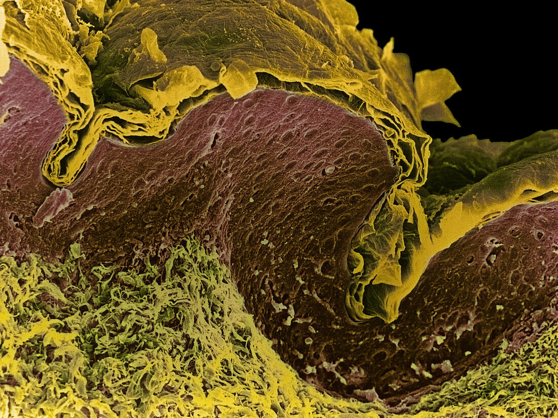
| Skin. Coloured scanning electron micrograph (SEM) of a section through human skin. At top is the stratum corneum (brown) of the epidermis,a cornified layer composed of flattened,dead skin cells which form the surface of the skin. The dead cells from this layer are continuously being shed and replaced from cells from the living epidermal layers below (purple). The lowest layer seen here is the dermis (yellow). This is a thick layer of fibrous connective tissue which supports and nourishes the epidermis. The skin is the body's largest organ,and accounts for around 15% of the weight of the body. Magnification unknown | |
| Licence : | Droits gérés |
| Crédit: | Science Photo Library / Gschmeissner, Steve |
| Taille de l’image : | 2640 px × 1981 px |
| Model Release : | Non requis |
| Property Release : | Non requis |
| Restrictions : | - |
Prix pour cette image À partir de 45 €
Produit vendu
(Calendrier, Carte postale, Carte de vœux, Impression sur textile, Packaging etc)
À partir de 45 €
Usage commercial
(Affichage, Annonce presse, Annonce TV, Carte, Digital - hors rés. sociaux, Digital - rés. sociaux etc)
À partir de 45 €
Éditorial
(Digital, Journal, Livre, Livre pratique, Magazine, Télévision etc)
À partir de 60 €
Usage non-commercial
(Digital - hors rés. sociaux, Digital - rés. sociaux etc)
À partir de 120 €