Sea urchin embryo
Numéro d’image : 11875505
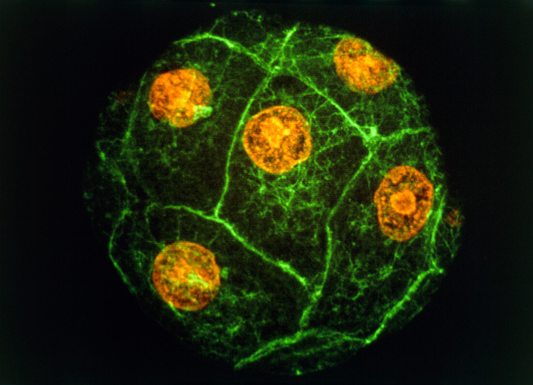
| Sea urchin embryo. Immunofluorescence micrograph of a sea urchin embryo at the 8-16 cell stage. The orange structures are the cell nuclei. The bound- aries between different cells show up as green lines. Following fertilization an embryo undergoes multiple rounds cell division,dividing each time into double the number of cells (1,2,4,8 ...). Eventually a hollow ball of hundreds of cells is produced,which then starts to differentiate. This picture was made by exposing the embryo to fluor- escent antibodies that bind to certain proteins in the cell. The embryo was then viewed with a laser- scanning light microscope,which makes the anti- bodies fluoresce. Magnification unknown | |
| Licence : | Droits gérés |
| Crédit: | Science Photo Library / PROF. G. SCHATTEN |
| Taille de l’image : | 5181 px × 3741 px |
| Model Release : | Non requis |
| Property Release : | Non requis |
| Restrictions : | - |
Prix pour cette image À partir de 45 €
Produit vendu
(Calendrier, Carte postale, Carte de vœux, Impression sur textile, Packaging etc)
À partir de 45 €
Usage commercial
(Affichage, Annonce presse, Annonce TV, Carte, Digital - hors rés. sociaux, Digital - rés. sociaux etc)
À partir de 45 €
Éditorial
(Digital, Journal, Livre, Livre pratique, Magazine, Télévision etc)
À partir de 60 €
Usage non-commercial
(Digital - hors rés. sociaux, Digital - rés. sociaux etc)
À partir de 120 €