Four-cell embryo
Numéro d’image : 11875144
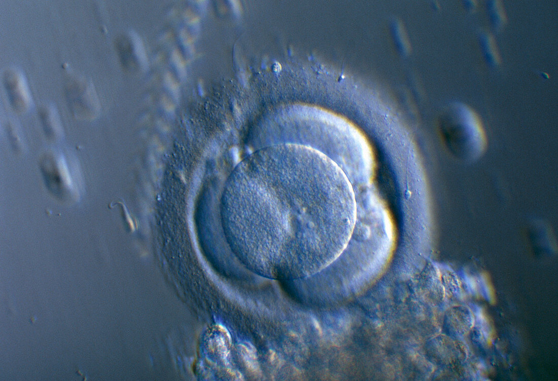
| Four-cell embryo. Light micrograph of a 4-cell human embryo. The cells,or blastomeres,result from two divisions of the fertilized egg. This stage is normally reached around 40 hours after fertilisation. The cells are surrounded by the zona pellucida layer. Outside this at lower right are cumulus corona cells (small circles),the remains of the corona radiata which supported and nourished the egg while it was in the ovary. Photographed at the Hospital Cochin,Paris,France. Magnification unknown | |
| Licence : | Droits gérés |
| Crédit: | Science Photo Library / Goetgheluck, Pascal |
| Taille de l’image : | 5184 px × 3543 px |
| Model Release : | Non requis |
| Property Release : | Non requis |
| Restrictions : | - |
Prix pour cette image À partir de 45 €
Produit vendu
(Calendrier, Carte postale, Carte de vœux, Impression sur textile, Packaging etc)
À partir de 45 €
Usage commercial
(Affichage, Annonce presse, Annonce TV, Carte, Digital - hors rés. sociaux, Digital - rés. sociaux etc)
À partir de 45 €
Éditorial
(Digital, Journal, Livre, Livre pratique, Magazine, Télévision etc)
À partir de 60 €
Usage non-commercial
(Digital - hors rés. sociaux, Digital - rés. sociaux etc)
À partir de 120 €
Mots clés
- 4 cellules,
- aucun,
- biologie,
- biologique,
- blastomère,
- corona radiata,
- corps humain,
- CUMULUS CORONA,
- de développement,
- développement,
- du développment,
- embryogenèse,
- embryologie,
- embryon,
- embryon humain,
- embryonnaire,
- en bonne santé,
- étape 4 cellules,
- humain,
- membrane pellucide,
- microscope optique,
- microscopie optique,
- multicellulaire,
- normal,
- personne,
- pluricellulaire,
- reproduction,
- sain,
- zona pellicida,
- zona pellucida,
- zone pellucide