Ultrasound scan of an IVF embryo in the uterus
Numéro d’image : 11875133
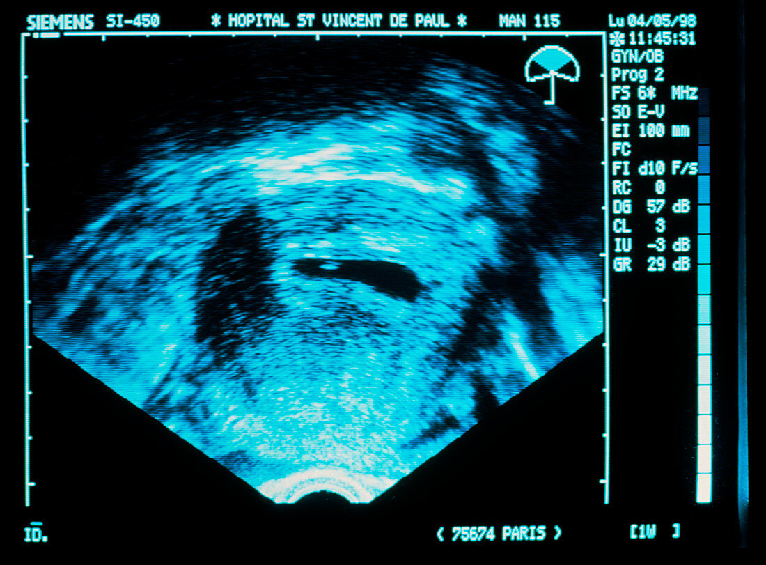
| Embryo ultrasound scan. Ultrasound scan of an early embryo conceived through in vitro fertilisation (IVF) implanted in the wall of the uterus. The uterine cavity is the dark oval at centre,with the tiny implanted embryo seen as the blue dot near its left edge. For IVF,sperm was introduced to the mother's egg in a laboratory. After fertilisation,the embryo was incubated for a few days before being implanted into the woman's uterus. This ultrasound scan was taken to ensure that implantation had occurred properly. Ultrasound uses high-frequency sound waves to build up a picture of the body's internal structures | |
| Licence : | Droits gérés |
| Crédit: | Science Photo Library / Goetgheluck, Pascal |
| Taille de l’image : | 5531 px × 4080 px |
| Model Release : | Non requis |
| Property Release : | Non requis |
| Restrictions : | - |
Prix pour cette image À partir de 45 €
Produit vendu
(Calendrier, Carte postale, Carte de vœux, Impression sur textile, Packaging etc)
À partir de 45 €
Usage commercial
(Affichage, Annonce presse, Annonce TV, Carte, Digital - hors rés. sociaux, Digital - rés. sociaux etc)
À partir de 45 €
Éditorial
(Digital, Journal, Livre, Livre pratique, Magazine, Télévision etc)
À partir de 60 €
Usage non-commercial
(Digital - hors rés. sociaux, Digital - rés. sociaux etc)
À partir de 120 €