Coloured SEM of blastocyst embryo hatching
Numéro d’image : 11875094
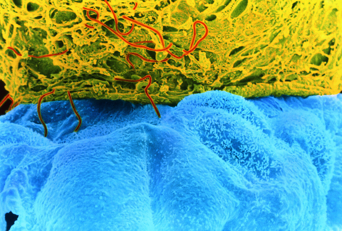
| Hatching blastocyst embryo. Coloured scanning electron micrograph (SEM) of a human embryo at the blastocyst stage,five days after fertilisation. The embryo (blue) is hatching from the zona pellucida (green),a protein shell that originally surrounded the unfertilised egg. Sperm tails (red) are also visible. A blastocyst is a hollow ball of cells with a fluid centre. Each cell is called a blastomere. Most of these embryo cells will form the placenta and membranes around the embryo,and only a small group (the inner mass) form the embryo proper. At this stage the blastocyst is preparing to implant on the wall of the uterus (womb). Magnification: x1030 at 5x7cm size. Magnification: x3700 at 10x6.5 ins size | |
| Licence : | Droits gérés |
| Crédit: | Science Photo Library / Nikas, Dr. Yorgos |
| Taille de l’image : | 5884 px × 3984 px |
| Model Release : | Non requis |
| Property Release : | Non requis |
| Restrictions : | - |
Prix pour cette image À partir de 45 €
Produit vendu
(Calendrier, Carte postale, Carte de vœux, Impression sur textile, Packaging etc)
À partir de 45 €
Usage commercial
(Affichage, Annonce presse, Annonce TV, Carte, Digital - hors rés. sociaux, Digital - rés. sociaux etc)
À partir de 45 €
Éditorial
(Digital, Journal, Livre, Livre pratique, Magazine, Télévision etc)
À partir de 60 €
Usage non-commercial
(Digital - hors rés. sociaux, Digital - rés. sociaux etc)
À partir de 120 €