Coloured SEM of 8-cell human embryo drilled open
Numéro d’image : 11875075
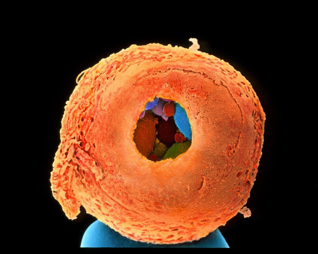
| Eight-cell embryo. Coloured scanning electron micrograph (SEM) of an 8-cell human embryo,three days old. It is drilled open to biopsy the embryo to test its genes for inherited diseases. The embryo is ontop of a pin (grey,at bottom). It is surrounded by a thick zona pellucida (brown) protective layer into which a window has been drilled. Large cells of the embryo are seen inside (coloured) while smaller spherical structures will degenerate. Embryo cells are called blastomeres & the embryo is termed a morula. It has yet to im- plant in the womb. By removing one or two blasto- meres,genes of the embryo can be examined. Magnification: x570 at 5x7cm size. x2,000 at 8x10ins | |
| Licence : | Droits gérés |
| Crédit: | Science Photo Library / Nikas, Dr. Yorgos |
| Taille de l’image : | 2660 px × 2130 px |
| Model Release : | Non requis |
| Property Release : | Non requis |
| Restrictions : | - |
Prix pour cette image À partir de 45 €
Produit vendu
(Calendrier, Carte postale, Carte de vœux, Impression sur textile, Packaging etc)
À partir de 45 €
Usage commercial
(Affichage, Annonce presse, Annonce TV, Carte, Digital - hors rés. sociaux, Digital - rés. sociaux etc)
À partir de 45 €
Éditorial
(Digital, Journal, Livre, Livre pratique, Magazine, Télévision etc)
À partir de 60 €
Usage non-commercial
(Digital - hors rés. sociaux, Digital - rés. sociaux etc)
À partir de 120 €