Coloured SEM of microvilli on surface of an embryo
Numéro d’image : 11875069
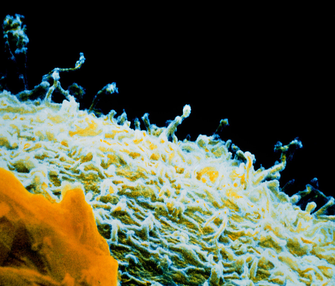
| Embryo surface. Coloured scanning electron micrograph of microvilli on the surface of an early embryo. The embryo is at the 2-4 cell stage,the egg has completed only one or two divisions after fertilization. The individual cells in the embryo are called blastomeres and it is a blastomere surface that is seen here. The surface is covered with numerous tiny projections called microvilli. These greatly increase the surface area of the cell and indicate the high metabolic activity of the cell. A probable fragment of a cumulus-corona cell is at bottom left (orange). Magnification: x4,200 at 6x7cm size | |
| Licence : | Droits gérés |
| Crédit: | Science Photo Library / PROFESSORS P.M. MOTTA & S. MAKABE |
| Taille de l’image : | 4843 px × 4140 px |
| Model Release : | Non requis |
| Property Release : | Non requis |
| Restrictions : | - |
Prix pour cette image À partir de 45 €
Produit vendu
(Calendrier, Carte postale, Carte de vœux, Impression sur textile, Packaging etc)
À partir de 45 €
Usage commercial
(Affichage, Annonce presse, Annonce TV, Carte, Digital - hors rés. sociaux, Digital - rés. sociaux etc)
À partir de 45 €
Éditorial
(Digital, Journal, Livre, Livre pratique, Magazine, Télévision etc)
À partir de 60 €
Usage non-commercial
(Digital - hors rés. sociaux, Digital - rés. sociaux etc)
À partir de 120 €