Coloured SEM of blastomere fragments in an embryo
Numéro d’image : 11875055
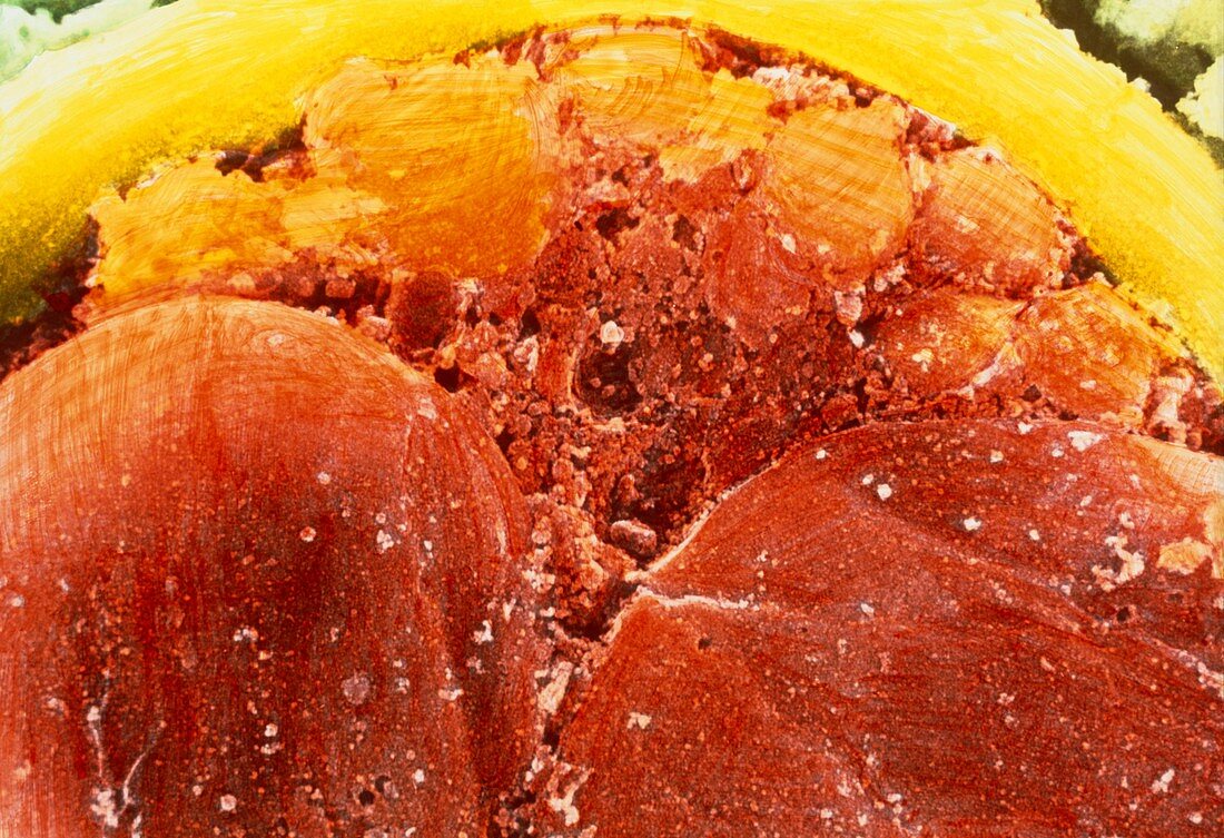
| 4-6 cell embryo. Coloured scanning electron micrograph of blastomeres and fragments in a 4-6 cell embryo. The blastomeres (large,orange,lower frame) are the cells formed from divisions of the fertilized egg (ovum). Smaller fragments of blastomeres are evident above them (upper frame). The production of small fragments such as these is quite normal in a rapidly dividing zygote. The fragments would be much larger and more numerous if the embryo were degenerating. Surrounding the embryo is a membranous envelope,the zona pellucida (yellow). Magnification: x2320 at 5x7cm size | |
| Licence : | Droits gérés |
| Crédit: | Science Photo Library / PROFESSORS P.M. MOTTA & S. MAKABE |
| Taille de l’image : | 5361 px × 3670 px |
| Model Release : | Non requis |
| Property Release : | Non requis |
| Restrictions : | - |
Prix pour cette image À partir de 45 €
Produit vendu
(Calendrier, Carte postale, Carte de vœux, Impression sur textile, Packaging etc)
À partir de 45 €
Usage commercial
(Affichage, Annonce presse, Annonce TV, Carte, Digital - hors rés. sociaux, Digital - rés. sociaux etc)
À partir de 45 €
Éditorial
(Digital, Journal, Livre, Livre pratique, Magazine, Télévision etc)
À partir de 60 €
Usage non-commercial
(Digital - hors rés. sociaux, Digital - rés. sociaux etc)
À partir de 120 €