Coloured SEM of an aborting 6-8 cell embryo
Numéro d’image : 11875046
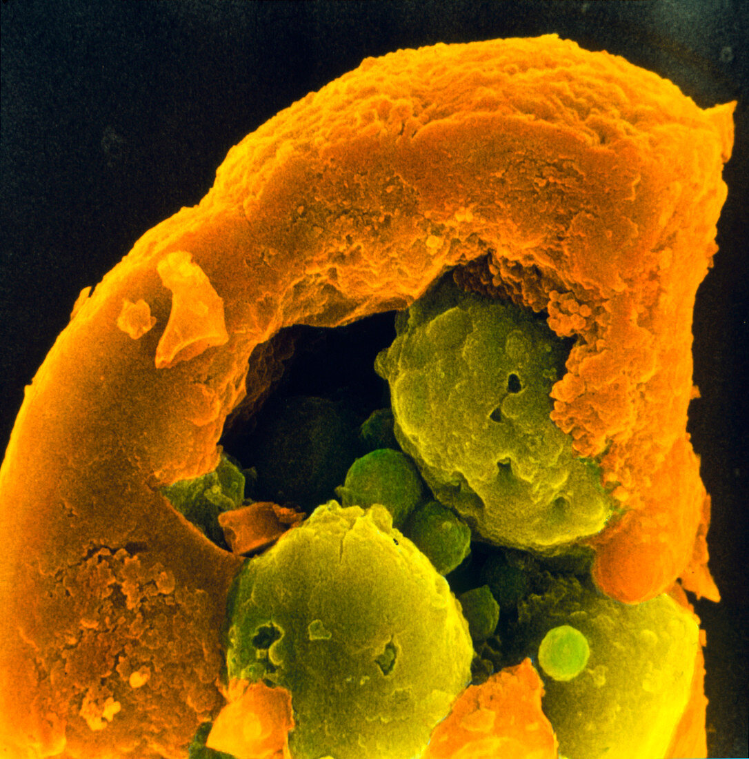
| Aborting embryo. Coloured scanning electron micrograph of an aborting 6-8 cell human embryo undergoing necrosis (tissue death). The blastomeres,the cells formed from divisions of the fertilized egg,are degenerating. Large blastomere cell fragments can be seen (green). The zona pellucida (orange),the membrane which surrounds the embryo,has become thick and compact. It has lost its typical striated appearance. Magnification: x2,000 at 6x6cm size | |
| Licence : | Droits gérés |
| Crédit: | Science Photo Library / PROFESSORS P.M. MOTTA & S. MAKABE |
| Taille de l’image : | 2868 px × 2904 px |
| Model Release : | Non requis |
| Property Release : | Non requis |
| Restrictions : | - |
Prix pour cette image À partir de 45 €
Produit vendu
(Calendrier, Carte postale, Carte de vœux, Impression sur textile, Packaging etc)
À partir de 45 €
Usage commercial
(Affichage, Annonce presse, Annonce TV, Carte, Digital - hors rés. sociaux, Digital - rés. sociaux etc)
À partir de 45 €
Éditorial
(Digital, Journal, Livre, Livre pratique, Magazine, Télévision etc)
À partir de 60 €
Usage non-commercial
(Digital - hors rés. sociaux, Digital - rés. sociaux etc)
À partir de 120 €