Coloured LM of a human embryo at two-cell stage
Numéro d’image : 11875007
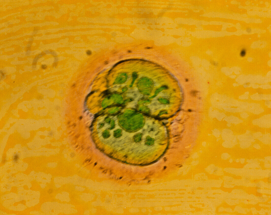
| Two-cell embryo. Coloured light micrograph of a human embryo at the 2-cell stage. It is seen 40 hours after fertilisation. The cells are coloured green,surrounded by the zona pellucida layer (orange). In the zona pellucida can be seen the heads (small black dots) of sperm. At this stage each cell of the embryo is called a blastomere. Inclusions (dark green) inside each cell are the cellular debris after cell division. The two-cell stage is a primitive embryo,following the first mitotic division. These cells will continue to divide to form a human foetus composed of millions of cells. Magnification: x200 at 6x7cm size. x275 at 4x5ins | |
| Licence : | Droits gérés |
| Crédit: | Science Photo Library / PROFESSOR P.M. MOTTA ET AL |
| Taille de l’image : | 3543 px × 2809 px |
| Model Release : | Non requis |
| Property Release : | Non requis |
| Restrictions : | - |
Prix pour cette image À partir de 45 €
Produit vendu
(Calendrier, Carte postale, Carte de vœux, Impression sur textile, Packaging etc)
À partir de 45 €
Usage commercial
(Affichage, Annonce presse, Annonce TV, Carte, Digital - hors rés. sociaux, Digital - rés. sociaux etc)
À partir de 45 €
Éditorial
(Digital, Journal, Livre, Livre pratique, Magazine, Télévision etc)
À partir de 60 €
Usage non-commercial
(Digital - hors rés. sociaux, Digital - rés. sociaux etc)
À partir de 120 €