SEM of a sectioned ovary at9-10 weeks of pregnancy
Numéro d’image : 11874991
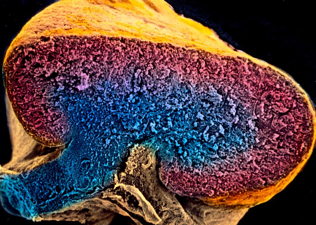
| Embryo. Coloured scanning electron micrograph of a sectioned human ovary of an embryo 9-10 weeks after fertilisation. The central part (blue) is called the medulla; it is surrounded by a cortical area (pale red) and by a surface epithelium (orange). Previously to this stage the medulla is occupied by clusters of primitive germ cells,known as sex cords,which have here disappeared and are replaced by a vascular stroma. The surface epithelium of the ovary gives rise in the 7th week to a second generation of sex cords,the cortical cords,which contain the final set of germ cells. Magnification: x170 at 6x7cm size | |
| Licence : | Droits gérés |
| Crédit: | Science Photo Library / PROFESSORS P.M. MOTTA & S. MAKABE |
| Taille de l’image : | 4979 px × 3543 px |
| Model Release : | Non requis |
| Property Release : | Non requis |
| Restrictions : | - |
Prix pour cette image À partir de 45 €
Produit vendu
(Calendrier, Carte postale, Carte de vœux, Impression sur textile, Packaging etc)
À partir de 45 €
Usage commercial
(Affichage, Annonce presse, Annonce TV, Carte, Digital - hors rés. sociaux, Digital - rés. sociaux etc)
À partir de 45 €
Éditorial
(Digital, Journal, Livre, Livre pratique, Magazine, Télévision etc)
À partir de 60 €
Usage non-commercial
(Digital - hors rés. sociaux, Digital - rés. sociaux etc)
À partir de 120 €