False-colour SEM of embryo at the morula stage
Numéro d’image : 11874965
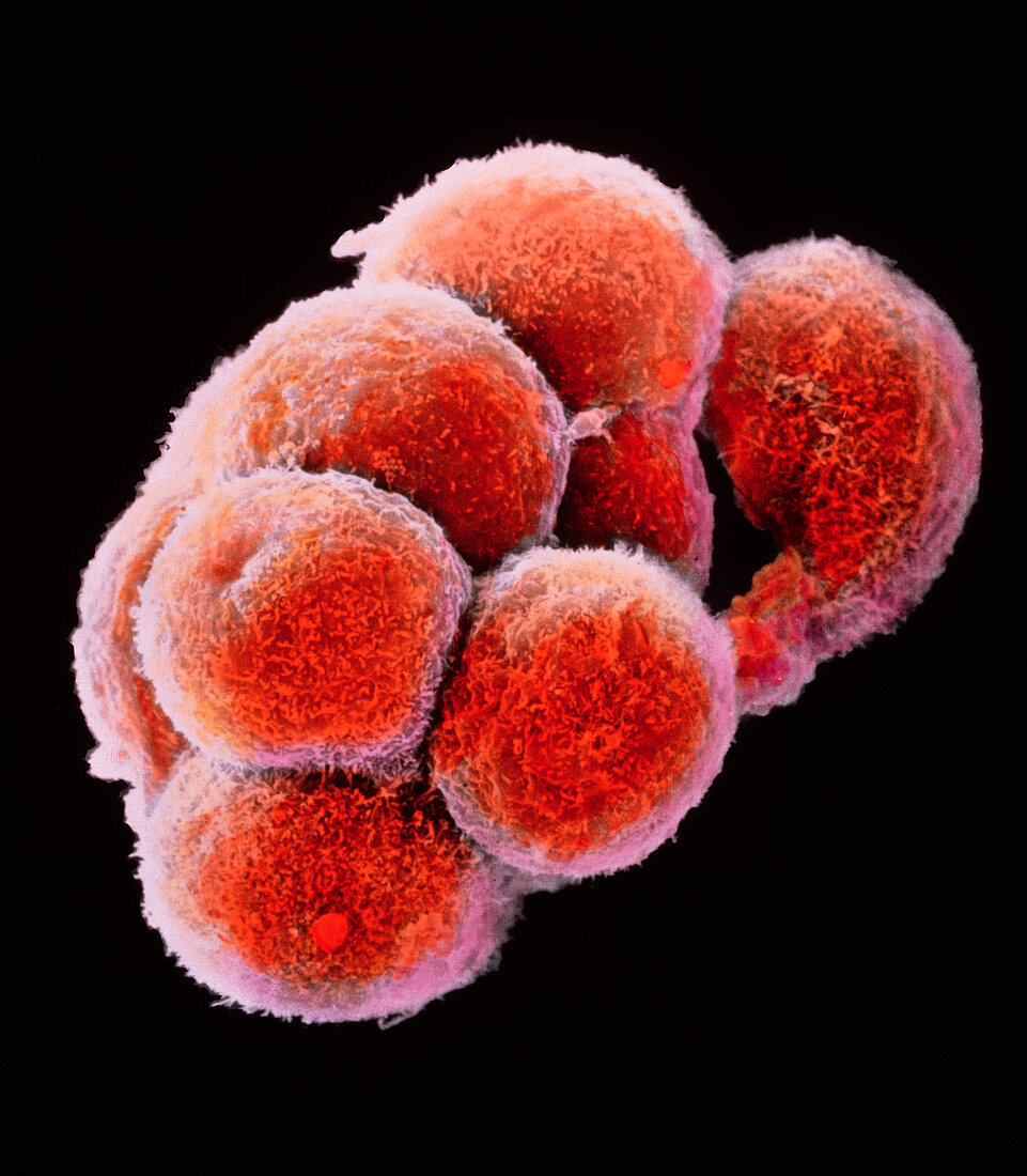
| Embryo development. False-colour scanning electron micrograph of an embryo at the early stage known as the morula. The egg reaches this phase about 4 days after fertilisation after a series of mitotic divisions. At this stage about 12-16 cells are present and are surrounded by a thin glycoprotein layer,the zona pellucida,which was here removed. The inner cells of the morula will give rise to the tissues of the embryo while the outer cells,covered here by microvilli (tiny orange ridges),will form the placenta. The morula will implant into the uterus six days after fertilisation. Magnification: x645 at 6x7cm size. Magnification: x1005 at 4x5 inch size. This is a mouse morula | |
| Licence : | Droits gérés |
| Crédit: | Science Photo Library / PROFESSORS P.M. MOTTA & J. VAN BLERKOM |
| Taille de l’image : | 3364 px × 3849 px |
| Model Release : | Non requis |
| Property Release : | Non requis |
| Restrictions : | - |
Prix pour cette image À partir de 45 €
Produit vendu
(Calendrier, Carte postale, Carte de vœux, Impression sur textile, Packaging etc)
À partir de 45 €
Usage commercial
(Affichage, Annonce presse, Annonce TV, Carte, Digital - hors rés. sociaux, Digital - rés. sociaux etc)
À partir de 45 €
Éditorial
(Digital, Journal, Livre, Livre pratique, Magazine, Télévision etc)
À partir de 60 €
Usage non-commercial
(Digital - hors rés. sociaux, Digital - rés. sociaux etc)
À partir de 120 €