False-colour TEM of 6-day-old fertilised ovum
Numéro d’image : 11874922
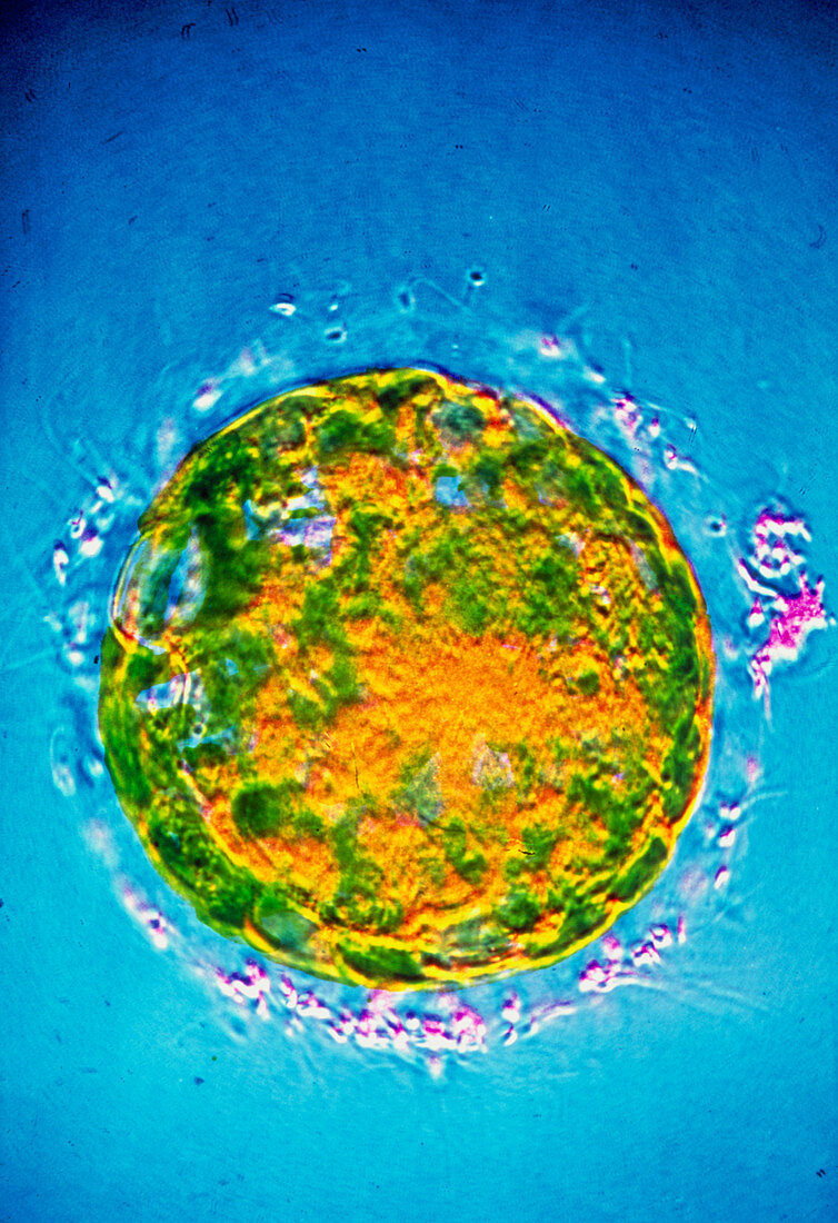
| False-colour transmission electron micrograph (TEM) of the human morula - a fertilised ovum - 6 days after fertilisation. The morula is an early stage of embryonic development; it is formed by successive cleavage of cells in the fertilised ovum. It consists of a solid ball of cells & may be considered as an intermediate stage between the zygote,the fertilised ovum before the advent of cleavage,& the blastocyst,the hollow ball of cells with an inner mass that attaches itself to the wall of the uterus. The ring of material (pink) around the edge of the morula is debris of sperm that failed to penetrate & fertilise the ovum. Magnification: x35 at 35mm size | |
| Licence : | Droits gérés |
| Crédit: | Science Photo Library / CNRI |
| Taille de l’image : | 2475 px × 3610 px |
| Model Release : | Non requis |
| Property Release : | Non requis |
| Restrictions : | - |
Prix pour cette image À partir de 45 €
Produit vendu
(Calendrier, Carte postale, Carte de vœux, Impression sur textile, Packaging etc)
À partir de 45 €
Usage commercial
(Affichage, Annonce presse, Annonce TV, Carte, Digital - hors rés. sociaux, Digital - rés. sociaux etc)
À partir de 45 €
Éditorial
(Digital, Journal, Livre, Livre pratique, Magazine, Télévision etc)
À partir de 60 €
Usage non-commercial
(Digital - hors rés. sociaux, Digital - rés. sociaux etc)
À partir de 120 €