Cell division
Numéro d’image : 11874829
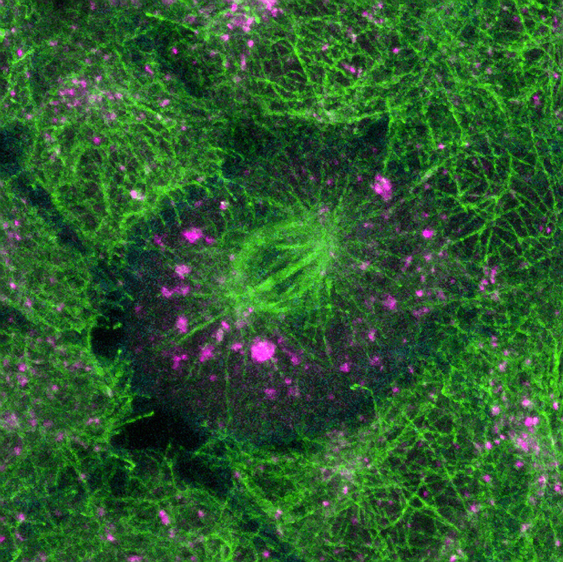
| Cell division. Confocal light micrograph of a dividing cell. The mitotic spindle (green,centre) functions to separate the split chromosomes (chromatids),moving one set into each daughter cell. The structure has been made visible by fluorescently-labelled antibodies to microtubulin,a protein of the spindle and cytoskeleton. Protein from the Golgi apparatus,a network of vesicles involved in processing and transporting cellular products,is labelled purple. A confocal microscope detects light only from the focal point of its objective lens,allowing images of thin sections of an intact specimen to be obtained. Magnification unknown | |
| Licence : | Droits gérés |
| Crédit: | Science Photo Library / Reichelt, Stefanie |
| Taille de l’image : | 3543 px × 3536 px |
| Model Release : | Non requis |
| Property Release : | Non requis |
| Restrictions : | - |
Prix pour cette image À partir de 45 €
Produit vendu
(Calendrier, Carte postale, Carte de vœux, Impression sur textile, Packaging etc)
À partir de 45 €
Usage commercial
(Affichage, Annonce presse, Annonce TV, Carte, Digital - hors rés. sociaux, Digital - rés. sociaux etc)
À partir de 45 €
Éditorial
(Digital, Journal, Livre, Livre pratique, Magazine, Télévision etc)
À partir de 60 €
Usage non-commercial
(Digital - hors rés. sociaux, Digital - rés. sociaux etc)
À partir de 120 €
Mots clés
- anaphase,
- animal,
- anticorps,
- axe,
- balayage au laser,
- broche,
- colorant,
- cytosquelette,
- division cellulaire,
- filaments,
- fluorescence,
- fluorescent,
- Golgi,
- immunofluorescence,
- marqué,
- metaphase,
- micrographe optique confocal,
- microscope optique,
- microscope optique confocal,
- microscopie optique,
- microtubule,
- microtubules,
- mitose,
- mitotique,
- protéines,
- scanning laser,
- tache,
- teindre,
- teinture,
- tige