Sea urchin cell division
Numéro d’image : 11874815
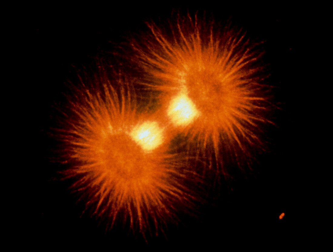
| Mitosis. Immunofluorescence micrograph of the "spindle" in a newly fertilized sea urchin embryo just before cell division. This cell,whose circu- lar outline can just be seen,is in the metaphase of mitosis (division of the nucleus). The spindle is the bright band in the middle,and consists of fibres called microtubules. The dark band in the middle of the spindle is where the chromosomes have lined up prior to being pulled apart into two identical sets. The bright bodies at lower left & upper right are the poles of the spindle,or cen- trioles. The rays of microtubules (asters) around these join the centrioles to the other parts of the cell. Magnification unknown | |
| Licence : | Droits gérés |
| Crédit: | Science Photo Library / PROF. G. SCHATTEN |
| Taille de l’image : | 4146 px × 3138 px |
| Model Release : | Non requis |
| Property Release : | Non requis |
| Restrictions : | - |
Prix pour cette image À partir de 45 €
Produit vendu
(Calendrier, Carte postale, Carte de vœux, Impression sur textile, Packaging etc)
À partir de 45 €
Usage commercial
(Affichage, Annonce presse, Annonce TV, Carte, Digital - hors rés. sociaux, Digital - rés. sociaux etc)
À partir de 45 €
Éditorial
(Digital, Journal, Livre, Livre pratique, Magazine, Télévision etc)
À partir de 60 €
Usage non-commercial
(Digital - hors rés. sociaux, Digital - rés. sociaux etc)
À partir de 120 €