Immunofluorescent micrograph of sea urchin mitosis
Numéro d’image : 11874814
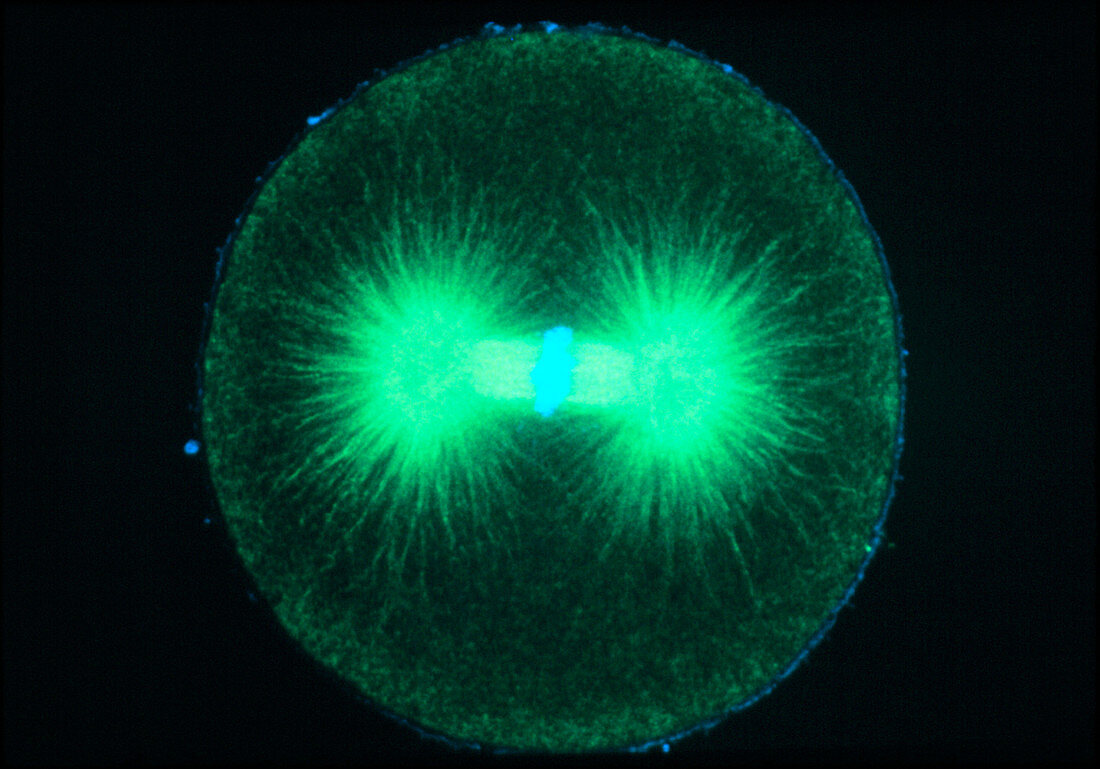
| Mitosis. Immunofluorescence micrograph of a newly fertilized sea urchin embryo during the metaphase of mitosis (division of the nucleus). This cell is about to divide into two. The chromosomes (blue) have aligned in a band across the middle of a "spindle" of fibres called microtubules (green). These will pull the chromosomes apart into two identical sets to form the nuclei of the daughter cells. The bright green bodies at left and right are the poles of the spindle,or centrioles. This picture was made by treating the cell with fluorescent antibodies that bind to specific proteins. The image was then captured by a laser- scanning light microscope. Magnification unknown | |
| Licence : | Droits gérés |
| Crédit: | Science Photo Library / PROF. G. SCHATTEN |
| Taille de l’image : | 3661 px × 2560 px |
| Model Release : | Non requis |
| Property Release : | Non requis |
| Restrictions : | - |
Prix pour cette image À partir de 45 €
Produit vendu
(Calendrier, Carte postale, Carte de vœux, Impression sur textile, Packaging etc)
À partir de 45 €
Usage commercial
(Affichage, Annonce presse, Annonce TV, Carte, Digital - hors rés. sociaux, Digital - rés. sociaux etc)
À partir de 45 €
Éditorial
(Digital, Journal, Livre, Livre pratique, Magazine, Télévision etc)
À partir de 60 €
Usage non-commercial
(Digital - hors rés. sociaux, Digital - rés. sociaux etc)
À partir de 120 €