Cell division
Numéro d’image : 11874759
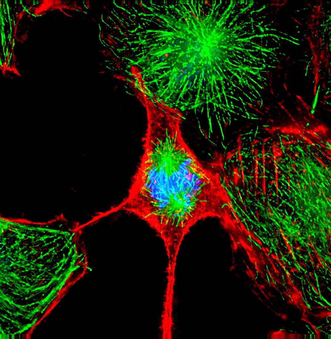
| Cell division. Immunofluorescent light micrograph of a cell during the metaphase stage of mitosis (cell division). It is surrounded by interphase (resting) cells. During metaphase,the DNA- containing chromosomes (blue) attach to the microtubules (green) and form a line (the metaphase plate) on the microtubule spindle. The microtubules pull the chromosomes apart towards either end of the cell. The cell will eventually split in two,forming genetically-identical daughter cells. Immunofluorescence uses antibodies to attach fluorescent dyes to specific cell tissues | |
| Licence : | Droits gérés |
| Crédit: | Science Photo Library / DR PAUL ANDREWS, UNIVERSITY OF DUNDEE |
| Taille de l’image : | 2923 px × 2992 px |
| Model Release : | Non requis |
| Property Release : | Non requis |
| Restrictions : | - |
Prix pour cette image À partir de 45 €
Produit vendu
(Calendrier, Carte postale, Carte de vœux, Impression sur textile, Packaging etc)
À partir de 45 €
Usage commercial
(Affichage, Annonce presse, Annonce TV, Carte, Digital - hors rés. sociaux, Digital - rés. sociaux etc)
À partir de 45 €
Éditorial
(Digital, Journal, Livre, Livre pratique, Magazine, Télévision etc)
À partir de 60 €
Usage non-commercial
(Digital - hors rés. sociaux, Digital - rés. sociaux etc)
À partir de 120 €
Mots clés
- A.D.N.,
- acide désoxyribonucléique,
- ADN,
- biologie,
- biologique,
- cellule,
- cycle,
- cyclique,
- cytologie,
- cytologique,
- diviser,
- division cellulaire,
- étape,
- génétique,
- immunofluorescence,
- interphase,
- metaphase,
- microscope optique,
- microscopie optique,
- microtubules,
- mitose,
- mitotique,
- phase,
- plaque de métaphase,
- plaque métaphasique,
- répétitif,
- réplication,
- répliquer,
- stade