Cell division
Numéro d’image : 11874757
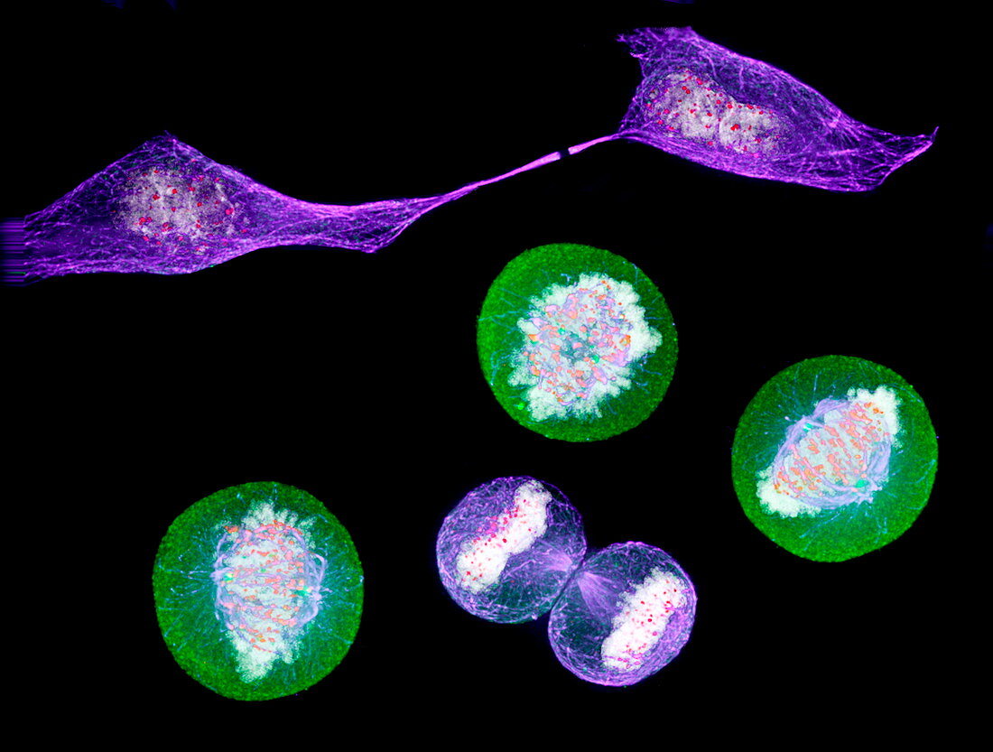
| Cell division. Immunofluorescent light micrograph of five HeLa cancer cells at different stages of cell division (mitosis). During mitosis,the genetic material of the cell divides and moves to opposite ends of the cell. The cell then splits into two daughter cells. Immunofluorescence uses antibodies to attach fluorescent dyes to specific cell tissues | |
| Licence : | Droits gérés |
| Crédit: | Science Photo Library / DR PAUL ANDREWS, UNIVERSITY OF DUNDEE |
| Taille de l’image : | 3398 px × 2572 px |
| Model Release : | Non requis |
| Property Release : | Non requis |
| Restrictions : | - |
Prix pour cette image À partir de 45 €
Produit vendu
(Calendrier, Carte postale, Carte de vœux, Impression sur textile, Packaging etc)
À partir de 45 €
Usage commercial
(Affichage, Annonce presse, Annonce TV, Carte, Digital - hors rés. sociaux, Digital - rés. sociaux etc)
À partir de 45 €
Éditorial
(Digital, Journal, Livre, Livre pratique, Magazine, Télévision etc)
À partir de 60 €
Usage non-commercial
(Digital - hors rés. sociaux, Digital - rés. sociaux etc)
À partir de 120 €
Mots clés
- anaphase,
- biologie,
- biologique,
- cancer,
- cancéreux,
- cellule,
- cellule HeLa,
- cellules,
- cinq,
- cycle,
- cyclique,
- cytokinèse,
- cytologie,
- cytologique,
- diviser,
- division cellulaire,
- génétique,
- immunofluorescence,
- interphase,
- metaphase,
- microscope optique,
- microscopie optique,
- microtubule,
- microtubules,
- mitose,
- mitotique,
- prophase,
- quintette,
- répétitif,
- réplication,
- répliquer,
- telophase