Cell division
Numéro d’image : 11874716
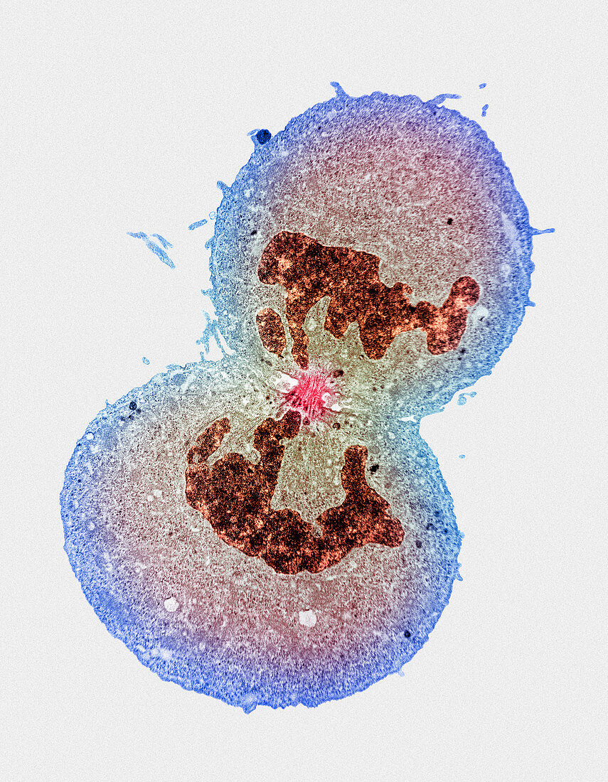
| Cell division. Coloured transmission electron micrograph of cytokinesis (division of a cell's cytoplasm) after mitosis of a tissue-cultured human embryonic kidney cell. In this late phase of cell division,telophase,the nucleus (brown) has divided in two. Each nucleus contains identical genetic material of the original mother cell. Microtubules and actin filaments (at centre,red) were the means of transporting the genetic material to the daughter cells. At telophase,the plasma membrane in the middle of the dividing cell is drawn in to form a cleavage furrow,which finally breaks the cell into two complete daughter cells. Magnification: x1800 at 6x7cm size | |
| Licence : | Droits gérés |
| Crédit: | Science Photo Library / Murti, Dr. Gopal |
| Taille de l’image : | 3312 px × 4259 px |
| Model Release : | Non requis |
| Property Release : | Non requis |
| Restrictions : | - |
Prix pour cette image À partir de 45 €
Produit vendu
(Calendrier, Carte postale, Carte de vœux, Impression sur textile, Packaging etc)
À partir de 45 €
Usage commercial
(Affichage, Annonce presse, Annonce TV, Carte, Digital - hors rés. sociaux, Digital - rés. sociaux etc)
À partir de 45 €
Éditorial
(Digital, Journal, Livre, Livre pratique, Magazine, Télévision etc)
À partir de 60 €
Usage non-commercial
(Digital - hors rés. sociaux, Digital - rés. sociaux etc)
À partir de 120 €
Mots clés
- agrandissement,
- cellules de rein,
- cellules rénales,
- corps humain,
- cytokinèse,
- cytologie,
- division cellulaire,
- foetal,
- foetale,
- foetus,
- histologie,
- humain,
- images,
- M.E.T.,
- MET,
- micrographie,
- microscope,
- microscope électronique à transmission,
- microtubules,
- mitose,
- photos au microscope,
- sujets,
- telophase