Col TEM of metaphase cell division of a HeLa cell
Numéro d’image : 11874701
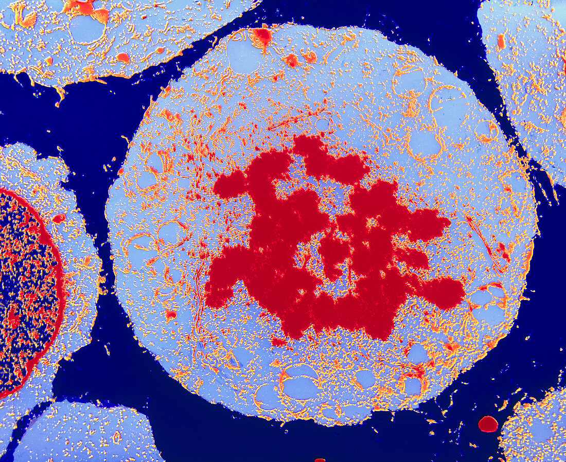
| Metaphase cell division. Coloured transmission electron micrograph of a section through a HeLa cell during metaphase cell division (mitosis). At centre are chromosomes (red); during metaphase the chromosomes line up in one plane in the centre of the cell,in preparation for nuclear division. The chromosomes are moved by spindle microtubules (not seen). At this stage of mitosis,the nuclear membrane has broken down. Organelles (white) are visible in the cytoplasm. During mitosis a cell produces two genetically identical daughter cells. HeLa cells are human cancer cells cultured in the laboratory and used in cancer research. Magnification: x2200 at 6x4.5cm size | |
| Licence : | Droits gérés |
| Crédit: | Science Photo Library / Murti, Dr. Gopal |
| Taille de l’image : | 4951 px × 4042 px |
| Model Release : | Non requis |
| Property Release : | Non requis |
| Restrictions : | - |
Prix pour cette image À partir de 45 €
Produit vendu
(Calendrier, Carte postale, Carte de vœux, Impression sur textile, Packaging etc)
À partir de 45 €
Usage commercial
(Affichage, Annonce presse, Annonce TV, Carte, Digital - hors rés. sociaux, Digital - rés. sociaux etc)
À partir de 45 €
Éditorial
(Digital, Journal, Livre, Livre pratique, Magazine, Télévision etc)
À partir de 60 €
Usage non-commercial
(Digital - hors rés. sociaux, Digital - rés. sociaux etc)
À partir de 120 €