Coloured LM of mature human oocyte
Numéro d’image : 11874226
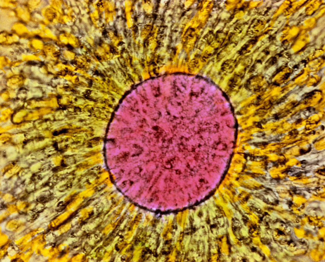
| Mature oocyte. Coloured light micrograph of a human secondary oocyte (mature egg). At centre is the rounded egg (pink) as seen in the ovary before ovulation. The egg cell undergoes meiotic division giving rise to two cells: a secondary oocyte and a polar body (not seen). The polar body degenerates and has no function. Here,directly around the egg is the zona pellucida single layer of cells (unfocused); outside the zona pellucida are several layers of cells making up the corona radiata (yellow). These cells support and nourish the developing egg in the ovary. Magnification: x560 at 6x7cm size. x740 at 4x5ins | |
| Licence : | Droits gérés |
| Crédit: | Science Photo Library / PROFESSOR P.M. MOTTA ET AL |
| Taille de l’image : | 5123 px × 4141 px |
| Model Release : | Non requis |
| Property Release : | Non requis |
| Restrictions : | - |
Prix pour cette image À partir de 45 €
Produit vendu
(Calendrier, Carte postale, Carte de vœux, Impression sur textile, Packaging etc)
À partir de 45 €
Usage commercial
(Affichage, Annonce presse, Annonce TV, Carte, Digital - hors rés. sociaux, Digital - rés. sociaux etc)
À partir de 45 €
Éditorial
(Digital, Journal, Livre, Livre pratique, Magazine, Télévision etc)
À partir de 60 €
Usage non-commercial
(Digital - hors rés. sociaux, Digital - rés. sociaux etc)
À partir de 120 €