False-colour SEM of an egg at ovulation
Numéro d’image : 11874212
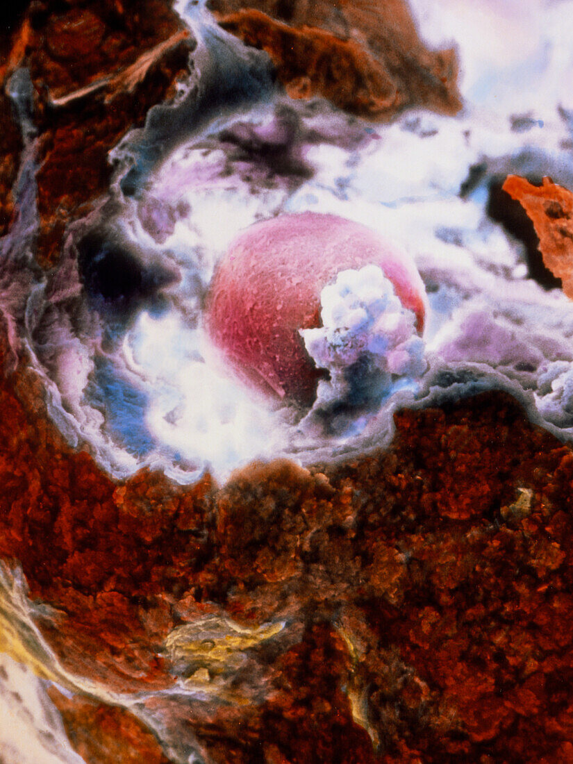
| Ovulation. False-colour scanning electron micrograph of the ovulation process. The egg (pink at centre) has ruptured the external surface of the ovary (brown),known as the germinal epithelium,and has started its journey through the Fallopian tube towards the uterus. The image clearly shows the thick glycoprotein layer (pink),the zona pellucida,which entirely surrounds the egg. The egg is also partly surrounded by cells (granulosa cells) and fluid (liquor folliculi),seen here in white and turquoise,which formerly provided nutrients and protection to the egg in the ovary. Magnification: x177 at 6x7cm size. Magnification: x255 at 4x5 inch size | |
| Licence : | Droits gérés |
| Crédit: | Science Photo Library / PROFESSORS P.M. MOTTA & J. VAN BLERKOM |
| Taille de l’image : | 2300 px × 3066 px |
| Model Release : | Non requis |
| Property Release : | Non requis |
| Restrictions : | - |
Prix pour cette image À partir de 45 €
Produit vendu
(Calendrier, Carte postale, Carte de vœux, Impression sur textile, Packaging etc)
À partir de 45 €
Usage commercial
(Affichage, Annonce presse, Annonce TV, Carte, Digital - hors rés. sociaux, Digital - rés. sociaux etc)
À partir de 45 €
Éditorial
(Digital, Journal, Livre, Livre pratique, Magazine, Télévision etc)
À partir de 60 €
Usage non-commercial
(Digital - hors rés. sociaux, Digital - rés. sociaux etc)
À partir de 120 €