False-colour SEM of a spermatozoon on uterus wall
Numéro d’image : 11874103
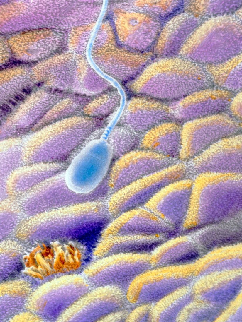
| A spermatozoon on the uterine mucosa. False-colour scanning electron micrograph of a spermatozoon floating over the endometrium,the internal wall of the uterus. Spermatozoa have pear-shaped heads about 7 microns long and a tail about 60 microns long. This image also shows the two different types of simple columnar cells which populate this area: ciliated (orange bottom left) and secretory (pink,yellow). During their journey through the uterus towards the Fallopian tubes spermatozoa undergo a capacitation process which enable them to penetrate and then fertilise an egg. Magnification: x2200 at 6x7cm size. x3465 at 4x5ins | |
| Licence : | Droits gérés |
| Crédit: | Science Photo Library / UNIVERSITY LA SAPIENZA, ROME / DEPT. OF ANATOMY / PROF. P. MOTTA |
| Taille de l’image : | 3502 px × 4670 px |
| Model Release : | Non requis |
| Property Release : | Non requis |
| Restrictions : | - |
Prix pour cette image À partir de 45 €
Produit vendu
(Calendrier, Carte postale, Carte de vœux, Impression sur textile, Packaging etc)
À partir de 45 €
Usage commercial
(Affichage, Annonce presse, Annonce TV, Carte, Digital - hors rés. sociaux, Digital - rés. sociaux etc)
À partir de 45 €
Éditorial
(Digital, Journal, Livre, Livre pratique, Magazine, Télévision etc)
À partir de 60 €
Usage non-commercial
(Digital - hors rés. sociaux, Digital - rés. sociaux etc)
À partir de 120 €