Colour SEM of microplicae on epithelium of cervix
Numéro d’image : 11873864
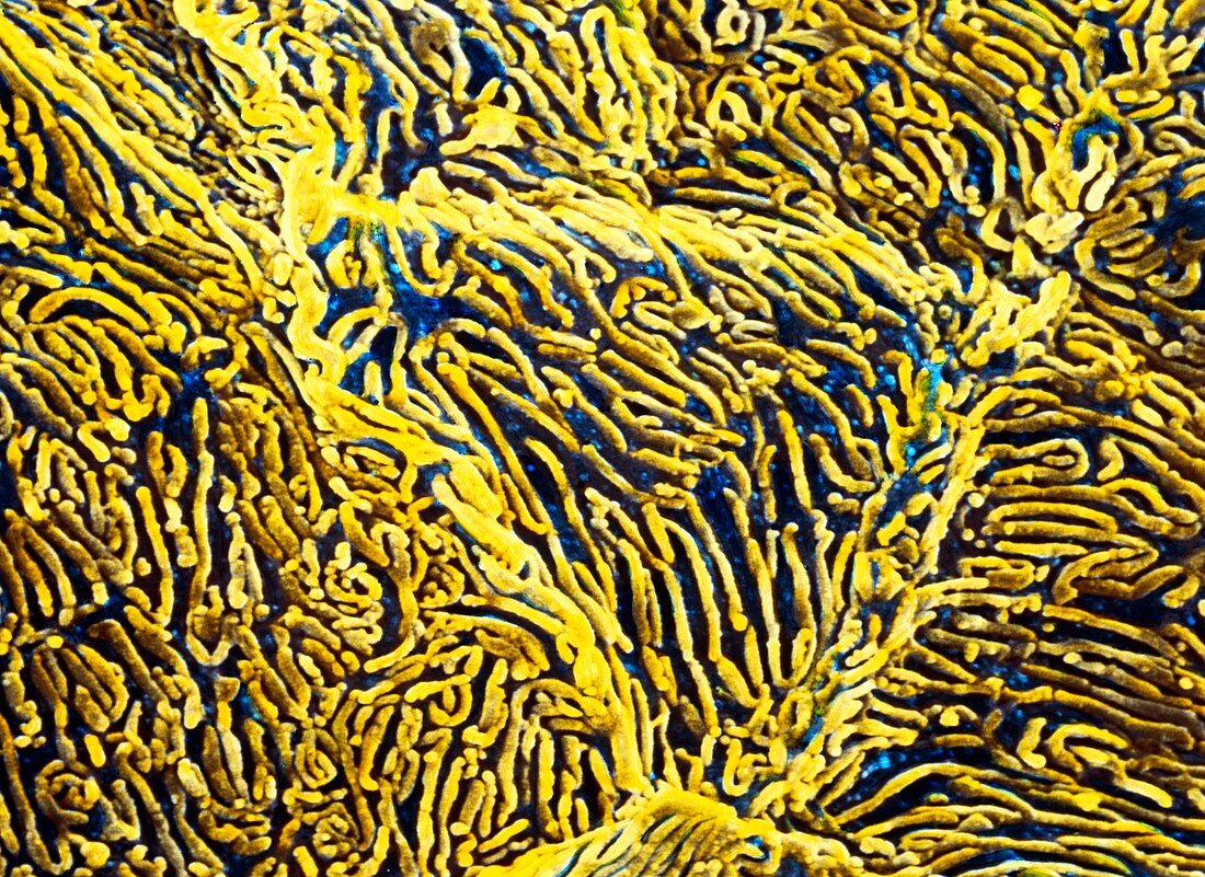
| Microplicae of vagina. Coloured scanning electron micrograph (SEM) of microplicae on the surface epithelium of the vaginal mucosa. The vagina is the muscular tube that forms the passageway between the cervix and vulva. Here,the surface of flattened squamous epithelial cells is shown (one cell is triangular at centre). These cells are covered in tiny,elongated folds called microplicae. Microplicae help to prevent the vaginal epithelium from drying out. They trap surface secretions (most notably mucus) within their folds,to keep the epithelium moist and lubricated. Magnification: x2800 at 5x7cm size | |
| Licence : | Droits gérés |
| Crédit: | Science Photo Library / PROFESSORS P.M. MOTTA & S. MAKABE |
| Taille de l’image : | 4949 px × 3602 px |
| Model Release : | Non requis |
| Property Release : | Non requis |
| Restrictions : | - |
Prix pour cette image À partir de 45 €
Produit vendu
(Calendrier, Carte postale, Carte de vœux, Impression sur textile, Packaging etc)
À partir de 45 €
Usage commercial
(Affichage, Annonce presse, Annonce TV, Carte, Digital - hors rés. sociaux, Digital - rés. sociaux etc)
À partir de 45 €
Éditorial
(Digital, Journal, Livre, Livre pratique, Magazine, Télévision etc)
À partir de 60 €
Usage non-commercial
(Digital - hors rés. sociaux, Digital - rés. sociaux etc)
À partir de 120 €