Coloured SEM of fallopian tube cilia & microvilli
Numéro d’image : 11873861
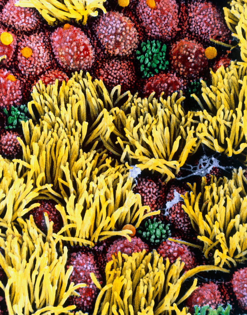
| Fallopian tube cells. Coloured high resolution scanning electron micrograph of cilia and micro- villi on the surface of the fallopian tube epithelium. Fallopian tubes are ducts that lead from the ovaries to the uterus. The epithelium consists of columnar cells,many of which have cilia (yellow). The cilia beat is towards the uterus,aiding transport of the egg from the ovary. Coloured red are the secretory cells with their microvilli projections. These cells secrete a substance (orange) that maintains a moist environment in the tube and may provide nutrients for the egg. Magnification: x2100 at 6x7cm size | |
| Licence : | Droits gérés |
| Crédit: | Science Photo Library / UNIVERSITY LA SAPIENZA, ROME / DEPT. OF ANATOMY / PROF. P. MOTTA |
| Taille de l’image : | 2814 px × 3582 px |
| Model Release : | Non requis |
| Property Release : | Non requis |
| Restrictions : | - |
Prix pour cette image À partir de 45 €
Produit vendu
(Calendrier, Carte postale, Carte de vœux, Impression sur textile, Packaging etc)
À partir de 45 €
Usage commercial
(Affichage, Annonce presse, Annonce TV, Carte, Digital - hors rés. sociaux, Digital - rés. sociaux etc)
À partir de 45 €
Éditorial
(Digital, Journal, Livre, Livre pratique, Magazine, Télévision etc)
À partir de 60 €
Usage non-commercial
(Digital - hors rés. sociaux, Digital - rés. sociaux etc)
À partir de 120 €