LM of a section through healthy cervix epithelium
Numéro d’image : 11873860
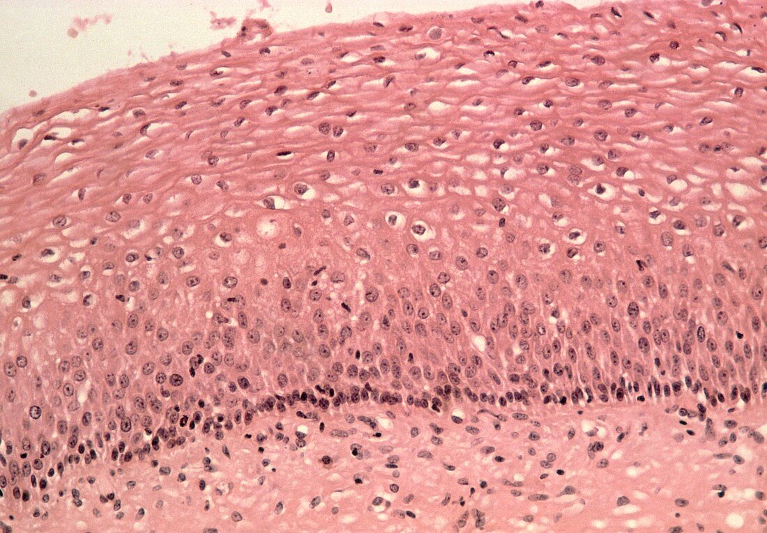
| Cervical epithelium. Light micrograph of a section through the epithelium of a biopsy taken from a healthy human cervix. The cervix is the narrow passage that connects the vagina and the uterus. The epithelial cells show a typically stratified (layered) form. Cell division is confined to a single layer of cuboidal cells (darkly stained layer,lower frame). As the cells mature they are pushed upwards and their nuclei (dark dots) thicken and eventually break down. The cell cytoplasm becomes progressively flattened until the cells are shed from the surface (top left). Haematoxylin and eosin stained. Magnification: x200 at 35mm size | |
| Licence : | Droits gérés |
| Crédit: | Science Photo Library / Walker, Dr. E. |
| Taille de l’image : | 5078 px × 3531 px |
| Model Release : | Non requis |
| Property Release : | Non requis |
| Restrictions : | - |
Prix pour cette image À partir de 45 €
Produit vendu
(Calendrier, Carte postale, Carte de vœux, Impression sur textile, Packaging etc)
À partir de 45 €
Usage commercial
(Affichage, Annonce presse, Annonce TV, Carte, Digital - hors rés. sociaux, Digital - rés. sociaux etc)
À partir de 45 €
Éditorial
(Digital, Journal, Livre, Livre pratique, Magazine, Télévision etc)
À partir de 60 €
Usage non-commercial
(Digital - hors rés. sociaux, Digital - rés. sociaux etc)
À partir de 120 €