Coloured SEM of a cut human umbilical cord & vein
Numéro d’image : 11873835
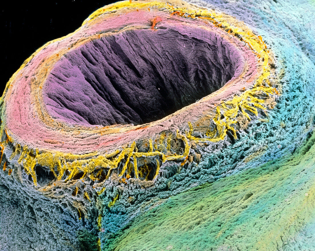
| Umbilical cord. Coloured Scanning Electron Micro- graph (SEM) of a sectioned human umbilical cord. Here,the cut umbilical vein (pink) reveals its inner folded lumen and a thin inner endothelial layer (purple). Most of the vein consists of several circular layers of smooth muscle. Around it is the jelly-like mucous connective tissue (blue) known as Wharton's jelly. Not seen are the two umbilical arteries which spiral around the vein. The umbilical cord is the attachment connecting the foetus with the placenta,and by which the foetus receives nourishment from the mother's blood. Magnification: x20 at 6x7cm size. x26 at 4x5ins | |
| Licence : | Droits gérés |
| Crédit: | Science Photo Library / PROFESSOR P.M. MOTTA & E. VIZZA |
| Taille de l’image : | 3543 px × 2830 px |
| Model Release : | Non requis |
| Property Release : | Non requis |
| Restrictions : | - |
Prix pour cette image À partir de 45 €
Produit vendu
(Calendrier, Carte postale, Carte de vœux, Impression sur textile, Packaging etc)
À partir de 45 €
Usage commercial
(Affichage, Annonce presse, Annonce TV, Carte, Digital - hors rés. sociaux, Digital - rés. sociaux etc)
À partir de 45 €
Éditorial
(Digital, Journal, Livre, Livre pratique, Magazine, Télévision etc)
À partir de 60 €
Usage non-commercial
(Digital - hors rés. sociaux, Digital - rés. sociaux etc)
À partir de 120 €