False-colour SEM of the Fallopian tube epithelium
Numéro d’image : 11873817
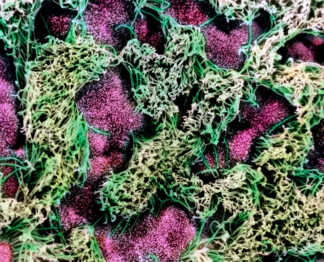
| Fallopian tube. False-colour scanning electron micrograph of the epithelium of the Fallopian tube (oviduct),the muscular tube connecting the ovary to the uterus. The image clearly shows the two different types of simple columnar cells which populate this area: ciliated (green) and secretory (soft purple). The beat of the ciliated cells facilitates the transport of the ovum from the ovary towards the uterus. The secretory cells are covered by a vast number of microvilli (tiny white dots) and secrete substances which maintain a moist environment and provide nutrients to the ovum. Magnification: x2230 at 6x7cm size. x2460 at 6x8cm | |
| Licence : | Droits gérés |
| Crédit: | Science Photo Library / UNIVERSITY LA SAPIENZA, ROME / DEPT. OF ANATOMY / PROF. P. MOTTA |
| Taille de l’image : | 4705 px × 3803 px |
| Model Release : | Non requis |
| Property Release : | Non requis |
| Restrictions : | - |
Prix pour cette image À partir de 45 €
Produit vendu
(Calendrier, Carte postale, Carte de vœux, Impression sur textile, Packaging etc)
À partir de 45 €
Usage commercial
(Affichage, Annonce presse, Annonce TV, Carte, Digital - hors rés. sociaux, Digital - rés. sociaux etc)
À partir de 45 €
Éditorial
(Digital, Journal, Livre, Livre pratique, Magazine, Télévision etc)
À partir de 60 €
Usage non-commercial
(Digital - hors rés. sociaux, Digital - rés. sociaux etc)
À partir de 120 €