False-colour SEM of uterus during pregnancy
Numéro d’image : 11873808
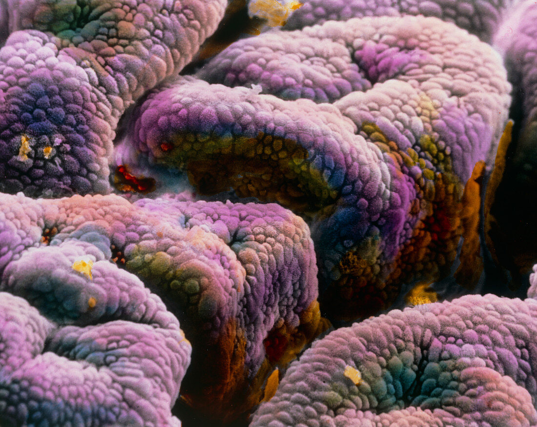
| The internal wall of the uterus in pregnancy. False-colour scanning electron micrograph of the uterine mucosa,the inner part of the endometrium,during pregnancy. The uterine mucosa is composed of simple columnar cells characterised by a vast number of microvilli on their surface. During pregnancy these cells increase their size and their secretory activity and the uterine mucosa shows a typical microfolded structure. The blood circulation in the underlying connective tissue also increases in order to provide more nutrient substances to the foetus. Magnification: x210 at 6x7cm size. Magnification: x330 at 4x5 inch size | |
| Licence : | Droits gérés |
| Crédit: | Science Photo Library / UNIVERSITY LA SAPIENZA, ROME / F. BARBERINI / PROFF. P. MOTTA |
| Taille de l’image : | 4843 px × 3854 px |
| Model Release : | Non requis |
| Property Release : | Non requis |
| Restrictions : | - |
Prix pour cette image À partir de 45 €
Produit vendu
(Calendrier, Carte postale, Carte de vœux, Impression sur textile, Packaging etc)
À partir de 45 €
Usage commercial
(Affichage, Annonce presse, Annonce TV, Carte, Digital - hors rés. sociaux, Digital - rés. sociaux etc)
À partir de 45 €
Éditorial
(Digital, Journal, Livre, Livre pratique, Magazine, Télévision etc)
À partir de 60 €
Usage non-commercial
(Digital - hors rés. sociaux, Digital - rés. sociaux etc)
À partir de 120 €