Light micrograph of normal human epididymis
Numéro d’image : 11873674
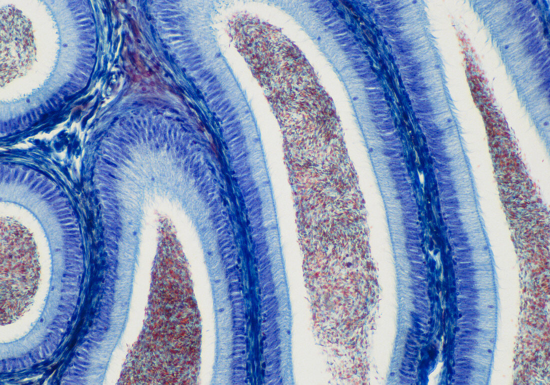
| Light micrograph of normal human epididymis. The bulk of the epididymis consists of a coiled duct seen here in oblique (right) and transverse (left) section. The lumen of the duct contains masses of spermatozoa (red). The clear space around these is crossed by sterocilia from the epithelial lining of the duct wall; these are in places seen as fine blue lines. The epithelium itself consists of a layer of columnar cells with elongated nuclei overlying a layer of rounded basal cells. The stratified dark blue layers between adjacent sections of duct are circularly arranged smooth muscle. Magnification: x50 at 35mm size | |
| Licence : | Droits gérés |
| Crédit: | Science Photo Library / Michler, Astrid & Hans-Frieder |
| Taille de l’image : | 5003 px × 3508 px |
| Model Release : | Non requis |
| Property Release : | Non requis |
| Restrictions : | - |
Prix pour cette image À partir de 45 €
Produit vendu
(Calendrier, Carte postale, Carte de vœux, Impression sur textile, Packaging etc)
À partir de 45 €
Usage commercial
(Affichage, Annonce presse, Annonce TV, Carte, Digital - hors rés. sociaux, Digital - rés. sociaux etc)
À partir de 45 €
Éditorial
(Digital, Journal, Livre, Livre pratique, Magazine, Télévision etc)
À partir de 60 €
Usage non-commercial
(Digital - hors rés. sociaux, Digital - rés. sociaux etc)
À partir de 120 €