Coloured CT scan showing the lungs and heart
Numéro d’image : 11873564
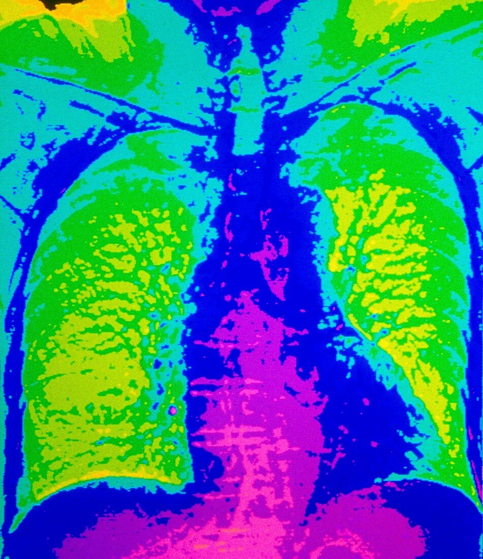
| Lungs and heart. Coloured Computed Tomography (CT) scan through the chest of a patient in front view,showing healthy lungs and heart. The lungs (green) are seen within the ribcage; lungs appear speckled here revealing their bronchial airways. The heart (at lower centre,pink/blue) is positioned between the lungs and lies partly inside the lung field at right. The diaphragm which separates the chest (thorax) from the abdomen is seen at lower frame. Spine bones run from the neck (at top) down the middle of the image. A CT scan is made using X- rays to produce "slice" images through the body | |
| Licence : | Droits gérés |
| Crédit: | Science Photo Library / Greim, John |
| Taille de l’image : | 3882 px × 4488 px |
| Model Release : | Non requis |
| Property Release : | Non requis |
| Restrictions : | - |
Prix pour cette image À partir de 45 €
Produit vendu
(Calendrier, Carte postale, Carte de vœux, Impression sur textile, Packaging etc)
À partir de 45 €
Usage commercial
(Affichage, Annonce presse, Annonce TV, Carte, Digital - hors rés. sociaux, Digital - rés. sociaux etc)
À partir de 45 €
Éditorial
(Digital, Journal, Livre, Livre pratique, Magazine, Télévision etc)
À partir de 60 €
Usage non-commercial
(Digital - hors rés. sociaux, Digital - rés. sociaux etc)
À partir de 120 €