SEM of lung alveoli
Numéro d’image : 11873557
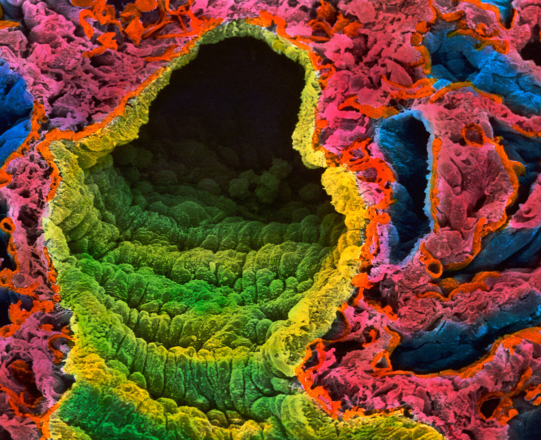
| Lung alveoli. Coloured Scanning Electron Micro- graph (SEM) of lung tissue,showing alveoli and a pulmonary blood vessel. At centre is the lumen of a large blood vessel (yellow) lined with elongated endothelial cells. To its right is an elongated blue arteriole; at upper left is a blue bronchio- lus with dark lumen. The other tissue (blue,pink) comprises alveoli air sacs with their interstital walls lined with capillaries. Bronchioli pass air into the alveoli where the gases of carbon dioxide and oxygen are exchanged during respiration. These gases pass directly out of,and into,the blood- stream. Magnification: x900 at 6x7cm size. Magnification: x1,150 at 4x5 inch size | |
| Licence : | Droits gérés |
| Crédit: | Science Photo Library / UNIVERSITY LA SAPIENZA, ROME / DEPT. OF ANATOMY / PROF. P. MOTTA |
| Taille de l’image : | 4063 px × 3304 px |
| Model Release : | Non requis |
| Property Release : | Non requis |
| Restrictions : | - |
Prix pour cette image À partir de 45 €
Produit vendu
(Calendrier, Carte postale, Carte de vœux, Impression sur textile, Packaging etc)
À partir de 45 €
Usage commercial
(Affichage, Annonce presse, Annonce TV, Carte, Digital - hors rés. sociaux, Digital - rés. sociaux etc)
À partir de 45 €
Éditorial
(Digital, Journal, Livre, Livre pratique, Magazine, Télévision etc)
À partir de 60 €
Usage non-commercial
(Digital - hors rés. sociaux, Digital - rés. sociaux etc)
À partir de 120 €