False-colour chest X-ray: normal 7 year-old child
Numéro d’image : 11873545
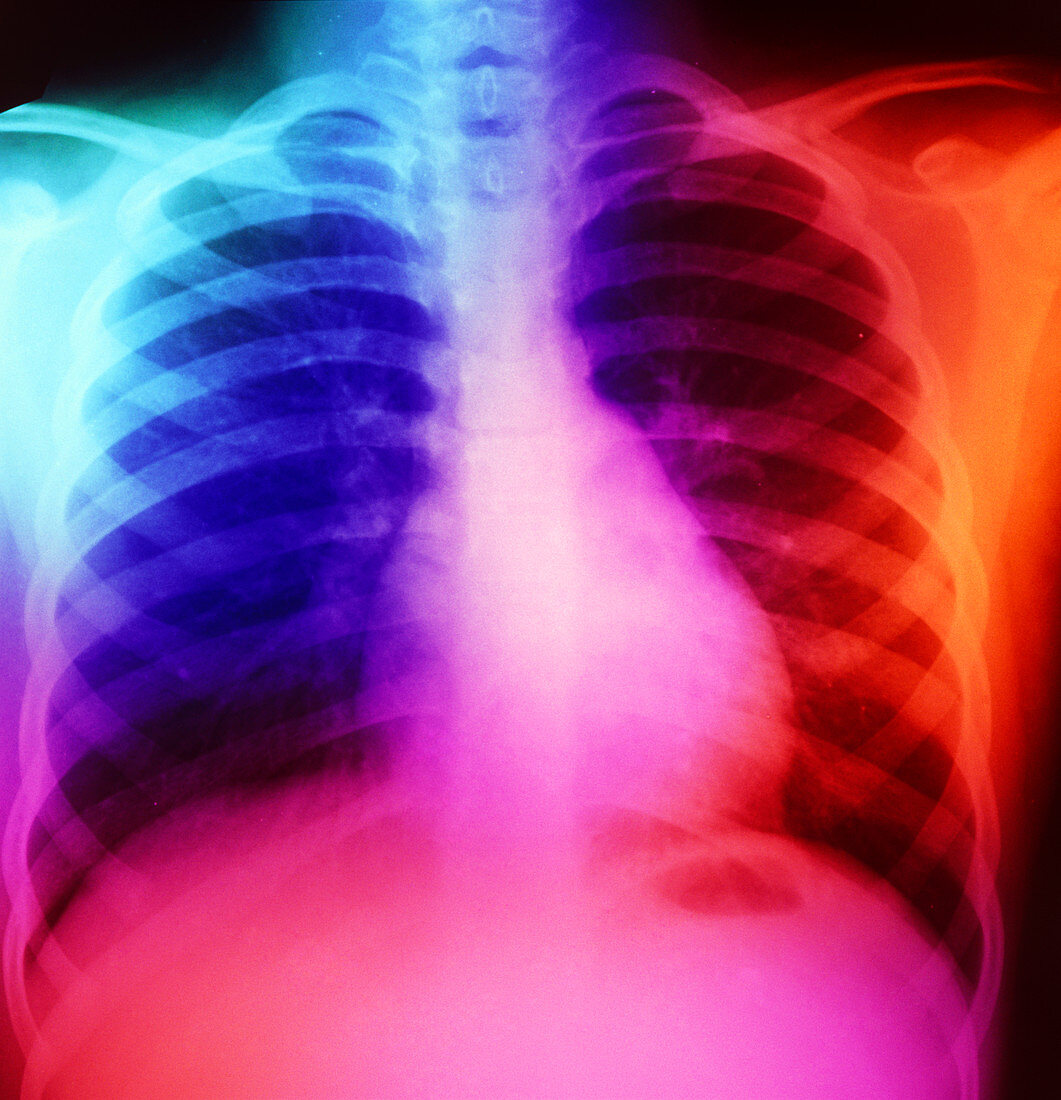
| False-colour normal chest X-ray of a 7 year old child. The image shows the skeleton of the spine,ribs and shoulder joints. Both lung fields appear dark,with opacities (pale areas) due to the bronchi & pulmonary blood vessels towards the mid- centre of each lung field. A normal-sized heart appears at centre-right in this posteroanterior (PA) image (that is,made with the radiographic film behind the patient's back & the X-ray source in front) | |
| Licence : | Droits gérés |
| Crédit: | Science Photo Library |
| Taille de l’image : | 4859 px × 5037 px |
| Model Release : | Non requis |
| Property Release : | Non requis |
| Restrictions : | - |
Prix pour cette image À partir de 45 €
Produit vendu
(Calendrier, Carte postale, Carte de vœux, Impression sur textile, Packaging etc)
À partir de 45 €
Usage commercial
(Affichage, Annonce presse, Annonce TV, Carte, Digital - hors rés. sociaux, Digital - rés. sociaux etc)
À partir de 45 €
Éditorial
(Digital, Journal, Livre, Livre pratique, Magazine, Télévision etc)
À partir de 60 €
Usage non-commercial
(Digital - hors rés. sociaux, Digital - rés. sociaux etc)
À partir de 120 €