False-colour SEM of a section of lung tissue
Numéro d’image : 11873537
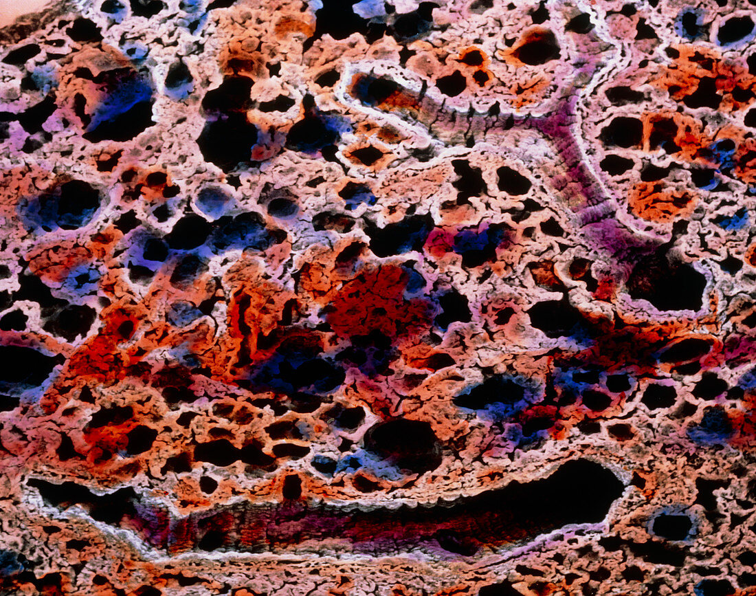
| Lung tissue. False-colour scanning electron micrograph of a section of lung tissue. The image shows many alveolar ducts (larger dark areas) together with many alveoli (smaller round dark areas). Two alveolar ducts are clearly visible (bottom centre and top right) as sectioned long tubules. The alveoli are separated by a porous membrane known as the interalveolar septum. The tiny openings in the interalveolar septum are sectioned capillaries and it is in this intricate network of capillaries that the exchange between carbon dioxide and oxygen takes place. Magnification: x70 at 6x7cm size. x110 at 4x5ins | |
| Licence : | Droits gérés |
| Crédit: | Science Photo Library / UNIVERSITY LA SAPIENZA, ROME / DEPT. OF ANATOMY / PROF. P. MOTTA |
| Taille de l’image : | 3739 px × 2948 px |
| Model Release : | Non requis |
| Property Release : | Non requis |
| Restrictions : | - |
Prix pour cette image À partir de 45 €
Produit vendu
(Calendrier, Carte postale, Carte de vœux, Impression sur textile, Packaging etc)
À partir de 45 €
Usage commercial
(Affichage, Annonce presse, Annonce TV, Carte, Digital - hors rés. sociaux, Digital - rés. sociaux etc)
À partir de 45 €
Éditorial
(Digital, Journal, Livre, Livre pratique, Magazine, Télévision etc)
À partir de 60 €
Usage non-commercial
(Digital - hors rés. sociaux, Digital - rés. sociaux etc)
À partir de 120 €